

Review
doi: 10.1016/j.str.2008.12.003.
Discovery of new GPCR biology: one receptor structure at a time
Affiliations
- PMID: 19141277
- PMCID: PMC2813843
- DOI: 10.1016/j.str.2008.12.003
Item in Clipboard
Review
Discovery of new GPCR biology: one receptor structure at a time
Structure.
.
Abstract
G-protein-coupled receptors (GPCRs) are the largest family of proteins in the human genome. Within the last year, we have witnessed a relative explosion in the amount of structural information available for the GPCR family with two new structures of opsin in the presence and absence of transducin peptide, four new structures of beta-adrenergic receptors, and a recent structure of the human adenosine A2A receptor. The new biological insight being gained, such as the highly divergent extracellular loops and areas of structural convergence within the transmembrane helices, allows us to chart a course for further investigation into this important class of membrane proteins.
Figures

Phylogenetic clustering of the class A (rhodopsin-like) GPCRs with the known structures mapped onto the appropriate cluster. The class A receptors may be divided into four groups termed α, β, γ and δ. The groups may be further divided according to the ligand binding characteristics of its members with the total number of receptors indicated in paranthesis. Here the α group is divided into amine, opsin, melatonin, prostaglandin and MECA (melatonin/EDG/cannabinoid/adenosine) receptors. The β group contains peptide receptors, the γ group contains the chemokine, melanin concentrating hormone (MCH) receptors and SOG (somatostatin/opioid/galanin) receptors. Finally, the δ group contains the MAS-related receptors, purine binding receptors and the glycoprotein receptors (Fredriksson et al., 2003). All GPCR structures have derived from the α group of receptors as shown.

Panel of representative GPCRs solved to date. Each group of receptors is represented by one structure all rendered with the same orientation and color scheme: transmembrane helices are colored light blue, intracellular regions are colored darker blue, extracellular regions are brown. Each ligand is colored orange and rendered as sticks, bound lipids are colored yellow and the conserved toggle switch tryptophan residue is rendered as spheres and colored green. This figure highlights the observed differences seen in the extracellular and intracellular domains as well as the small differences seen in the ligand binding orientations.
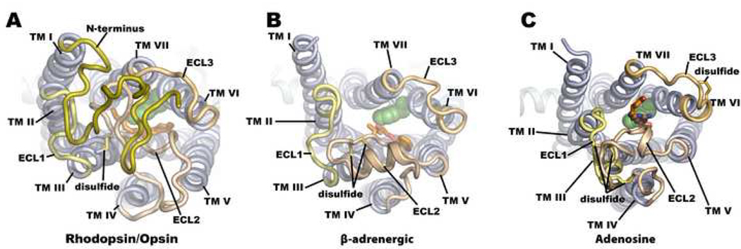
Extracellular view of A. rhodopsin B. β-adrenergic and C. adenosine A2A receptors. The ligand in each case is rendered as sticks and colored orange and the toggle switch tryptophan is rendered as van der Waals spheres and colored green. The extracellular domain in rhodopsin serves to occlude the retinal binding pocket, whereas in both the β-adrenergic and adenosine A2A receptors the extracellular domain is highly constrained and held away from the ligand binding pocket opening.

Ligand binding pocket of A. rhodopsin B. β-adrenergic and C. adenosine A2A receptors. Residues interacting with the ligand in each case (orange sticks) are colored as green sticks. Polar interactions where applicable are shown as yellow lines between the interacting atoms. A. The retinal binding pocket relies mainly on hydrophobic interactions in addition to a covalent linkage with TM VII. B. The β-adrenergic ligands interact with the receptors through two clusters of polar interactions. The first cluster is shown at the tail of the ligand carazolol where the positively charged secondary amine group and β-OH group participate in polar interactions with a conserved glutamate on TM III and asparagine on TM VII. The second grouping of polar interactions is with the head group of the ligand and a cluster of serine residues on TM V. C. The adenosine ligand ZM241385 forms mainly polar interactions between a primary amine group and an asparagine residue on TM VI and a glutamate on ECL2. A π-stacking interaction between the ligands heterocyclic group and a phenylalanine residue also on ECL2 plays a role in binding affinity.

Panel of intracellular interactions across the family of solved class A GPCRs. Bovine rhodopsin is the only receptor with an intact ionic lock interaction between the E/DRY motif and glutamate on TM VI. However, in the opsin structures the ionic lock is broken and the helical section of TM V is extended considerably relative to the inactive bovine rhodopsin. Human β2AR has a similar length TM V as bovine opsin, turkey β1AR and human A2A adenosine receptors, all of which have a disrupted ionic lock. With the exception of opsin and rhodopsin the DRY motif interacts with ICL2 through a polar interaction between the aspartate residue and either a serine or tyrosine residue on ICL2.
References
-
- Caffrey M. A lipid's eye view of membrane protein crystallization in mesophases. Curr Opin Struct Biol. 2000;10:486–497. - PubMed
-
- Caffrey M. Membrane protein crystallization. J Struct Biol. 2003;142:108–132. - PubMed
-
- Cherezov V, Clogston J, Papiz MZ, Caffrey M. Room to move: crystallizing membrane proteins in swollen lipidic mesophases. J Mol Biol. 2006;357:1605–1618. - PubMed
Publication types
MeSH terms
Substances
Grants and funding
LinkOut - more resources
Full Text Sources
