. 2009 Mar 15;385(2):358-67.
doi: 10.1016/j.virol.2008.12.014. Epub 2009 Jan 11.

Role of regulatory T cells in coronavirus-induced acute encephalitis

Affiliations

Role of regulatory T cells in coronavirus-induced acute encephalitis

Daniela Anghelina et al. Virology. .

Abstract

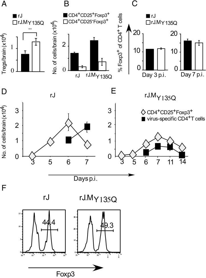
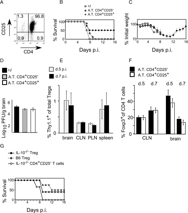
C57BL/6 mice infected with mouse hepatitis virus, strain JHM (JHMV) develop a rapidly fatal acute encephalitis. Previously, we showed that this disease is partially CD4 T cell-mediated since infection with a recombinant JHMV (rJ) mutated in only a single immunodominant CD4 T cell epitope (epitope M133, rJ.M(Y135Q)) results in a nonlethal disease. Increased mortality correlated with a greater number of JHMV-specific CD4 T cells in the brains of rJ compared to rJ.M(Y135Q)-infected mice. Here, we extend these results to show that the diminished number of virus-specific T cells correlates with a reduced cytokine/chemokine response in the infected brain. We also show that regulatory CD4 T cells (Tregs) are critical for mild disease in rJ.M(Y135Q)-infected mice because their depletion results in increased mortality. Further, a relative paucity of Tregs characterizes lethal infection because adoptive transfer of Tregs into rJ-infected mice increases survival from 0% to 50%. These results support the notion that clinical disease in coronavirus-induced acute encephalitis results from a balance between factors critical for virus clearance, such as virus-specific effector T cells and anti-inflammatory elements, such as Tregs. These findings also show that unlike chronic infections, in which an excessive number of Tregs contributes to pathogen persistence, Tregs in the setting of acute encephalitis may help to limit immunopathological disease without delaying virus clearance.


Figures

Fig. 1
Fig. 1
Regulatory T cells in the brains of mice inoculated with rJ or rJ.MY135Q. (A) Cells were harvested at 7 days p.i. from the brains of mice infected with rJ or rJ.MY135Q and stained for CD4, CD25 and Foxp3. The number of CD4+CD25+ cells that express Foxp3 is shown (n = 8 for each virus). ⁎⁎p < 0.02. (B) Numbers of CD4 T cells that are CD25+Foxp3+ or CD25Foxp3+ in brains of mice infected with rJ or rJ.MY135Q at 7 days p.i. (n = 5 for each virus). (C) Frequency of CD4 T cells that are Foxp3+ in CLN of rJ and rJ.MY135Q-infected mice (n = 3–5 mice for each virus at each time point). (D, E) Cells were harvested from the brains of mice infected with rJ or rJ.MY135Q and analyzed for numbers of Tregs and virus-specific CD4 T cells (epitopes M133, S358, S333-specific) by IFN-γ intracellular staining at the indicated times. Numbers of virus-specific CD4 T cells and Tregs are shown. Three to eight mice per virus were analyzed at each time point. (F) Cells were harvested from the brains of mice infected with rJ or rJ.MY135Q at 3 days p.i. and stained for CD4, CD25 and Foxp3. Percentage of CD4+CD25+ T cells expressing Foxp3 is indicated. Representative histograms are shown. Four mice infected with each virus were analyzed. Of note, very few CD4 T cells (approximately 2000) were detected in infected mice at day 3 p.i.
Fig. 2
Fig. 2
Functional and phenotypic characterization of Tregs harvested from infected mice. (A) Mononuclear cells harvested at 7 days p.i. from the brains of B6 mice infected with rJ or rJ.MY135Q were stained directly ex vivo for CD4, CD25, Foxp3 and GITR, CD103, CD69, CD62L or CD44. Cells harvested from a rJ.MY135Q-infected mouse are shown in the figure. (B) CD4+CD25+ (Tregs) and CD4+CD25 (non-Tregs) T cells were sorted from the CLN of rJ.MY135Q-infected B6 mice (7 days p.i.) as described in Materials and methods. CD4+CD25 and CD8+ cells were prepared from the lymph nodes of naive mice, labeled with CFSE, stimulated with 1 μg/ml anti-CD3 and incubated for 72 h with the sorted Tregs or non-Tregs and irradiated splenocytes at the indicated ratios (Treg or non-Treg: CD8 or CD4+CD25). Cells were analyzed for CFSE dilution (proliferation) by flow cytometry as described in Materials and methods.
Fig. 3
Fig. 3
Identification of epitope M133-specific Tregs in rJ-infected mice. (A) Cells were harvested from the brains of infected Foxp3-GFP mice and assayed for epitope M133-specific CD4 T cells by staining with specific and non-specific (human CLIP-specific) MHC class II tetramers and anti-CD4 antibody. Cells shown in the figure were gated for CD4 expression. (B) Frequencies of M133-specific Tregs (CD4+Foxp3+) and M133-specific non-Tregs (CD4+Foxp3) within total CD4 T cells in the brain are shown. Ten mice were analyzed.
Fig. 4
Fig. 4
Increased mortality and weight loss in rJ.MY135Q-infected mice after Treg depletion. (A) Mice were treated with 0.5 mg anti-CD25 antibody (mAb PC61) or rat IgG 3 days prior to infection with rJ.MY135Q. Percentage of Foxp3+CD25+ of total CD4 T cells in the brain at day 5 p.i. (8 days post treatment) is shown. (B, D) B6 mice were infected with rJ.MY135Q three days after treatment with anti-CD25 antibody (n = 10) or rat IgG (n =15) and monitored for survival (B) and weight loss (D). Results from three independent experiments are shown. Mortality (p < 0.05) and weight loss (p < 0.05 at days 8–13) were significantly increased in anti-CD25 mAb-treated mice. (C) Viral titers in the brain at day 5 p.i after treatment with mAb PC61 or rat IgG (n = 5 for each antibody).
Fig. 5
Fig. 5
Increased survival after adoptive transfer of Tregs into rJ-infected mice. (A) CD4+CD25+ T cells isolated from naive B6 spleens as described in Materials and methods were 95–98% pure as assessed by flow cytometry. (B) 3.7 × 105 CD4+CD25+ T cells were adoptively transferred into mice at day 1 after infection with rJ. As a control, rJ-infected mice received CD4+CD25 cells or PBS. Mice were monitored for survival (B) and weight loss (C). Three independent experiments were performed, n = 10 mice/group. Mice receiving CD4+CD25+ T cells survived at a higher frequency than those receiving CD4+CD25 cells or PBS (p < 0.05). (D) Virus titers were the same in rJ-infected recipients of CD4+CD25+ or CD4+CD25 T cells or PBS at day 7 p.i. Four mice were analyzed in each group. (E) Tregs were purified from the spleens of naive Thy1.1 mice and transferred into Thy1.2 B6 mice 1 day after infection with rJ. Mice were sacrificed at day 5 or 7 p.i. and Tregs from brains, spleens, CLN and peripheral (PLN) lymph nodes were analyzed for Thy1.1/1.2 expression. A total of five mice were analyzed at each day p.i. in two independent experiments. (F) Brains and CLN were harvested at days 5 and 7 p.i. from rJ-infected mice that received CD4+CD25+ or CD4+CD25 T cells. Foxp3+ CD4 T cells were determined by flow cytometry. (G) IL-10−/− (n = 5) or B6 Tregs (n = 4) or CD4+ CD25IL-10−/− T cells (n = 3) were transferred into B6 mice at day 1 p.i. Two independent experiments were performed. Mice were monitored for survival. A.T.—adoptive transfer.

References

    1. Anghelina D., Pewe L., Perlman S. Pathogenic role for virus-specific CD4 T cells in mice with coronavirus-induced acute encephalitis. Am. J. Pathol. 2006;169(1):209–222. - PMC - PubMed
    1. Belkaid Y. Regulatory T cells and infection: a dangerous necessity. Nat. Rev., Immunol. 2007;7(11):875–888. - PubMed
    1. Belkaid Y., Rouse B.T. Natural regulatory T cells in infectious disease. Nat. Immunol. 2005;6(4):353–360. - PubMed
    1. Bergmann C.C., Lane T.E., Stohlman S.A. Coronavirus infection of the central nervous system: host-virus stand-off. Nat. Rev., Microbiol. 2006;4(2):121–132. - PMC - PubMed
    1. Couper K.N., Blount D.G., Riley E.M. IL-10: the master regulator of immunity to infection. J. Immunol. 2008;180(9):5771–5777. - PubMed

Publication types