. 2008 Dec 15;22(24):3449-60.
doi: 10.1101/gad.1697808.

CPEB regulation of human cellular senescence, energy metabolism, and p53 mRNA translation

Affiliations

CPEB regulation of human cellular senescence, energy metabolism, and p53 mRNA translation

David M Burns et al. Genes Dev. .

Abstract

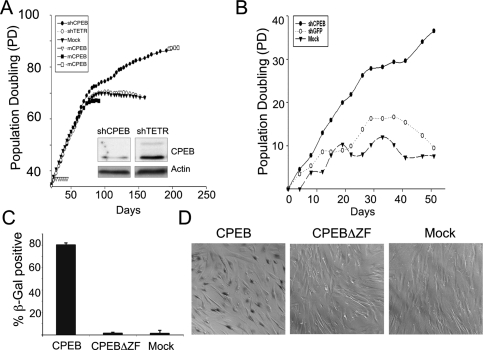
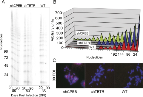
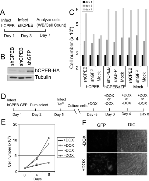
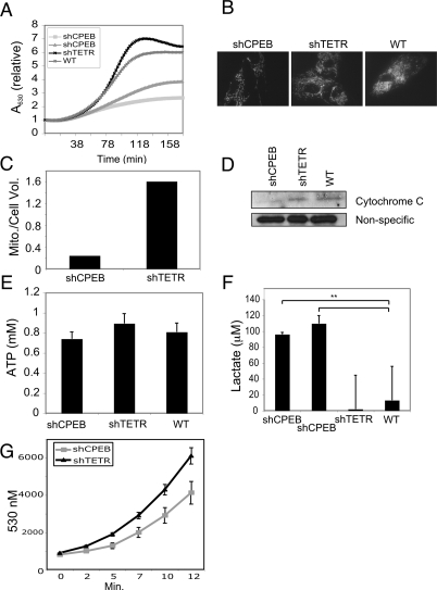
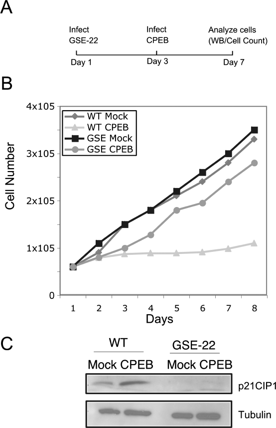
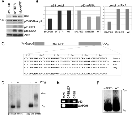
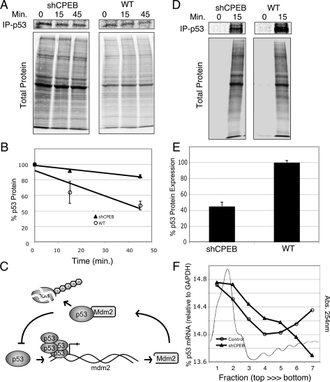
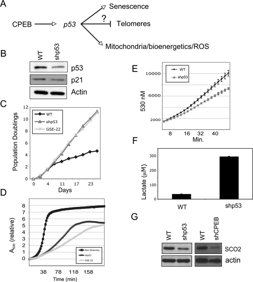
Cytoplasmic polyadenylation element-binding protein (CPEB) stimulates polyadenylation and translation in germ cells and neurons. Here, we show that CPEB-regulated translation is essential for the senescence of human diploid fibroblasts. Knockdown of CPEB causes skin and lung cells to bypass the M1 crisis stage of senescence; reintroduction of CPEB into the knockdown cells restores a senescence-like phenotype. Knockdown cells that have bypassed senescence undergo little telomere erosion. Surprisingly, knockdown of exogenous CPEB that induced a senescence-like phenotype results in the resumption of cell growth. CPEB knockdown cells have fewer mitochondria than wild-type cells and resemble transformed cells by having reduced respiration and reactive oxygen species (ROS), normal ATP levels, and enhanced rates of glycolysis. p53 mRNA contains cytoplasmic polyadenylation elements in its 3' untranslated region (UTR), which promote polyadenylation. In CPEB knockdown cells, p53 mRNA has an abnormally short poly(A) tail and a reduced translational efficiency, resulting in an approximately 50% decrease in p53 protein levels. An shRNA-directed reduction in p53 protein by about 50% also results in extended cellular life span, reduced respiration and ROS, and increased glycolysis. Together, these results suggest that CPEB controls senescence and bioenergetics in human cells at least in part by modulating p53 mRNA polyadenylation-induced translation.


Figures

Figure 1.
Figure 1.
CPEB is necessary for cellular senescence. (A) Human foreskin fibroblasts were infected with a lentiviruses encoding shRNA targeting CPEB mRNA (shCPEB), or the tetracycline resistance mRNA (shTETR), as a control. Additional cells were mock infected with an empty lentivirus. Population doublings were then determined by counting cells with a hemocytometer. Some cells were also infected with a retrovirus expressing mouse CPEB (because of differences in the primary sequence, mouse CPEB mRNA is not a target of the shCPEB), followed by cell counting. The inset shows a Western blot probed for CPEB and actin; these extracts were prepared from cells 40 d post-lentivirus infection. (B) Human lung fibroblasts (WI-38) were infected with lentiviruses targeting CPEB or, as a control GFP; mock refers to infection with an empty virus. The cells were counted as in A. (C,D) Foreskin fibroblasts previously infected with shCPEB were also infected with retroviruses expressing wild-type CPEB or a CPEB lacking a zinc finger, which renders the protein incapable of RNA binding. Some cells were also mock infected. The cells were then stained for b-galactosidase at acidic pH and were counted (C), and visualized by bright-field microscopy 3 d after retrovirus infection (D).
Figure 2.
Figure 2.
CPEB knockdown cells retain long telomeres. (A) DNA was extracted from skin fibroblasts infected with shCPEB and shTETR-containing lentiviruses 20, 50, or 90 d post-infection and used to determine telomere length by the T-OLA. (B) The relative telomere lengths derived from the analysis in A were quantified by scanning desitometry. (C) shCPEB, shTETR, and noninfected wild-type (WT) cells were fixed at 90 d post-infection and used for FISH to telomeric regions (telo-FISH) using a Cy3-conjugated locked nucleic acid oligonucleotide (LNA). The DNA was stained with DAPI.
Figure 3.
Figure 3.
CPEB-induced senescence-like phenotype is reversible. (A) Schematic representation of one sequence of events used to examine the reversibility of the CPEB-induced senescence-like phenotype. (B) Western blot for CPEB and tubulin derived from fibroblasts that were sequentially infected with a retrovirus encoding HA-tagged human CPEB (hCPEB-HA) followed by a lentivirus encoding shCPEB (two different sequences) or shGFP. (C) Cell numbers were determined following sequential infection of the viruses noted in A. (D) Schematic representation of the experimental timeline when cells were infected with a retrovirus expressing CPEB under the control of the tetracycline repressor. Skin fibroblasts were infected with a virus expressing a CPEB-GFP fusion protein under the control of the tetracycline response element (TET-ON), followed the next day by puromycin selection. Three days later, the cells were infected with a virus encoding the tetracycline repressor (TETR); the cells were then cultured for several days in the presence of DOX, which allows GFP-CPEB expression to remain high. Some cells were then cultured in medium lacking DOX, which will repress GFP-CPEB transcription. (E) Growth chart of two cell lines containing exogenous CPEB-GFP under control of the TETr cultured in the absence or presence of DOX. (F) Live cell images of GFP-CPEB containing cells cultured in the absence or presence of DOX.
Figure 4.
Figure 4.
Reduced respiration and mitochondrial number in CPEB knockdown cells. (A) Fibroblasts infected with shCPEB (targeted to two different sequences), shTETR, or empty virus (mock) were cultured for ∼47 d when they were used to measure oxygen consumption. (B) Z-plane stacks of confocal images obtained from live cells, some of which were infected with shCPEB or shTETR, stained with MitotrackerRed to visualize mitochondria. (C) Quantification of MitotrackerRed fluorescence from B. (D) Immunoblot of cytochrome C from wild-type, shTETR-, and shCPEB-infected cells. A nonspecific immuno-reactive band served as a loading control. (E) Wild-type, shCPEB, and shTETR cells were used to measure ATP concentration. (F) Lactate, an indicator of glycolysis, was determined in shCPEB (targeting two different sequences) and control cells. (G) Determination of relative ROS levels in cells expressing shCPEB or shTETR.
Figure 5.
Figure 5.
CPEB induced senescence requires p53. (A) Fibroblasts were infected with a retrovirus expressing GSE-22, a p53 dominant-negative peptide. Two days later, the cells were infected with a virus harboring CPEB; examination of the cells began on day 7. (B) Growth curves of GSE-22 or CPEB-infected or mock-infected cells. (C) Western blot analysis of p21 and tubulin in infected or mock-infected cells.
Figure 6.
Figure 6.
CPEB promotes cytoplasmic polyadenylation of p53 mRNA. (A) Extracts from shCPEB, shTETR, or mock-infected cells were probed with antibodies specific for p53, K382 acetylated p53, p21CIP1, p16INK4A, and actin. (B) The amount of p53 protein in A was quantified by densitometry, and the amount of p53 RNA in other infected cells was quantified by qRT–PCR; the ratios of these values were then plotted. (C) Diagram of the salient features of the p53 mRNA 3′ UTRs from several mammals; the CPEs and AAUAAA hexanucleotide are in bold. (Bottom) Sequence denotes dinucleotide substitutions in each of the two CPEs. (D) The human wild-type and mutated p53 3′ UTRs were radiolabeled and injected into Xenopus oocytes, which were then treated with progesterone to induce meiotic maturation and cytoplasmic polyadenylation. (E) Fibroblasts infected with a retrovirus encoding HA-CPEB or HA-CPEB lacking the zinc finger were used for HA antibody coimmunoprecipitation and RT–PCR detection of p53 mRNA and the non-CPE-containing GAPDH mRNA. (F) Ligation-mediated polyadenylation test (LM-PAT) assay was used to estimate the poly(A) tail length of p53 mRNA in wild-type and shCPEB knockdown cells.
Figure 7.
Figure 7.
CPEB controls p53 mRNA translation. (A,B) Wild-type and shCPEB-infected cells were incubated in methoinine and cysteine-free medium for 30 min, followed by an incubation with 35S-methionine and 35S-cysteine for 30 min, and then an incubation of up to 45 min with excess radioinert methionine and cysteine. p53 was immunoprecipitated from the extracts at 0, 15, and 45 min of the amino acid chase and resolved by SDS-PAGE, as was total cell protein. (C) Diagram illustrating a feedback loop where high levels of p53 induce transcription of the E3 ligase mdm2, which in turn induces p53 destruction. Ub refers to ubiquitin. (D,E) wild-type and shCPEB-infected cells were incubated in methionine and cysteine-free media for 1 h followed by incubation in 35S-methionine and 35S-cysteine for 15 min. p53 was then immunoprecipitated and analyzed by SDS-PAGE and phosphorimaging. The quantification of p53 levels in three different experiments is shown. (F) Extracts from wild-type and shCPEB-infected fibroblasts were centrifuged through 15%–50% sucrose gradients, fractionated, and scanned with 254-nm light. The relative amounts of p53 and GAPDH mRNAs were quantified by qRT–PCR. A representative polysome profile (absorbance at 254-nm light) from wild-type cells is shown.
Figure 8.
Figure 8.
p53 regulation of senescence and bioenergetics. (A) Proposed pathway in which CPEB, at least in part, influences senescence, telomere maintenance, and bioenergetics. (B) Western blot showing a shRNA-directed ∼50% knockdown of p53, which inhibits the expression of p21. (C) Growth curves of wild-type cells or cells infected with shp53 or GSE-22. (D) Oxygen consumption in cells infected with a nonsilencing shRNA, shp53 RNA, or GSE-22, a p53 inhibitory peptide. (E) Relative ROS levels in wild-type or shp53-infected cells. (F) Levels of lactate in wild-type and shp53 knockdown cells. (G) Western blot of SCO2 in CPEB and p53 knockdown cells.

References

    1. Archer H., Bar-Sagi D. Ras and Rac as activators of reactive oxygen species (ROS) Methods Mol. Biol. 2002;189:67–73. - PubMed
    1. Barnard D.C., Ryan K., Manley J.L., Richter J.D. Symplekin and xGLD-2 are required for CPEB-mediated cytoplasmic polyadenylation. Cell. 2004;119:641–651. - PubMed
    1. Barnard D.C., Cao Q., Richter J.D. Differential phosphorylation controls Maskin association with eukaryotic translation initiation factor 4E and localization on the mitotic apparatus. Mol. Cell. Biol. 2005;25:7605–7615. - PMC - PubMed
    1. Beausejour C.M., Krtolica A., Galimi F., Narita M., Lowe S.W., Yaswen P., Campisi J. Reversal of human cellular senescence: Roles of the p53 and p16 pathways. EMBO J. 2003;22:4212–4222. - PMC - PubMed
    1. Ben-Porath I., Weinberg R.A. The signals and pathways activating cellular senescence. Int. J. Biochem. Cell Biol. 2005;37:961–976. - PubMed

Publication types

MeSH terms

Substances