. 2008 Dec 15;22(24):3489-96.
doi: 10.1101/gad.1742908.

Cas6 is an endoribonuclease that generates guide RNAs for invader defense in prokaryotes

Affiliations

Cas6 is an endoribonuclease that generates guide RNAs for invader defense in prokaryotes

Jason Carte et al. Genes Dev. .

Abstract

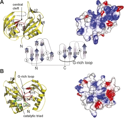
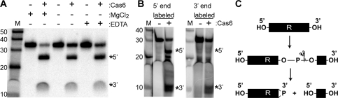
An RNA-based gene silencing pathway that protects bacteria and archaea from viruses and other genome invaders is hypothesized to arise from guide RNAs encoded by CRISPR loci and proteins encoded by the cas genes. CRISPR loci contain multiple short invader-derived sequences separated by short repeats. The presence of virus-specific sequences within CRISPR loci of prokaryotic genomes confers resistance against corresponding viruses. The CRISPR loci are transcribed as long RNAs that must be processed to smaller guide RNAs. Here we identified Pyrococcus furiosus Cas6 as a novel endoribonuclease that cleaves CRISPR RNAs within the repeat sequences to release individual invader targeting RNAs. Cas6 interacts with a specific sequence motif in the 5' region of the CRISPR repeat element and cleaves at a defined site within the 3' region of the repeat. The 1.8 angstrom crystal structure of the enzyme reveals two ferredoxin-like folds that are also found in other RNA-binding proteins. The predicted active site of the enzyme is similar to that of tRNA splicing endonucleases, and concordantly, Cas6 activity is metal-independent. cas6 is one of the most widely distributed CRISPR-associated genes. Our findings indicate that Cas6 functions in the generation of CRISPR-derived guide RNAs in numerous bacteria and archaea.


Figures

Figure 1.
Figure 1.
Cas6 is an endoribonuclease that cleaves CRISPR RNAs within repeat sequences. (A) psiRNA biogenesis pathway model. The primary CRISPR transcript contains unique invader targeting or guide sequences (colored blocks) flanked by direct repeat sequences (R). Cas6 catalyzes site-specific cleavage within each repeat, releasing individual invader targeting units. The Cas6 cleavage products undergo further processing to generate smaller mature psiRNA species. (B) Purified recombinant PfCas6 expressed in E. coli. The sizes (in kilodaltons) of protein markers (M) are indicated. (C) Radiolabeled RNAs (repeat–guide–repeat [R–g–R] or repeat alone [R], as diagrammed) were either uniformly or 5′-end-labeled and incubated in the absence (−) or presence (+) of PfCas6 protein (500 nM). Products were resolved by denaturing gel electrophoresis and visualized using a phosphorimager. The main cleavage products are indicated by a star or asterisk on the gel and in the diagram.
Figure 2.
Figure 2.
PfCas6 cleavage of a CRISPR RNA containing two repeat-guide RNA units. A uniformly radiolabeled substrate RNA containing two guide (invader targeting) sequences (yellow and green), two repeats (R) and a short (natural) 5′ leader (L) sequence was incubated with 1 μM PfCas6 protein and samples were analyzed by denaturing gel electrophoresis at the indicated times. The expected sizes and compositions of the RNA products (based on site-specific cleavage within each repeat) are indicated, as are the sizes of the marker RNAs (M).
Figure 3.
Figure 3.
Identification of the site of PfCas6 cleavage within the CRISPR repeat RNA. (A) The site of PfCas6 cleavage within the CRISPR repeat RNA was mapped by incubating 5′ end labeled repeat RNA with PfCas6 nuclease and comparing the size of the 5′ RNA cleavage product (arrow) with RNAse T1 (T1) and alkaline hydrolysis (OH) sequence ladders. (B) Potential secondary structure of P. furiosus repeat RNA with cleavage site indicated. (C) Analysis of cleavage of wild-type and cleavage site mutant (AA to GG) repeat RNAs with increasing concentrations (0, 1, 50, 200, and 500 nM) of PfCas6. (D) Native gel mobility shift analysis of wild-type and mutant repeat RNAs with increasing concentrations of PfCas6. The positions of the free (RNA) and protein-bound (RNP) RNAs are indicated. 5′ and 3′ cleavage products are indicated in both C and D. The sizes of RNA markers (M) are indicated in A and C.
Figure 4.
Figure 4.
CRISPR repeat sequence requirements for PfCas6 binding. (A) Detailed analysis of binding with a series of CRISPR-derived RNAs and mutants. The left panel illustrates the RNAs tested, with repeat (R) and invader targeting (yellow blocks) sequences, and PfCas6 cleavage site (dashed lines) indicated. Blue block denotes an insertion, dashed block denotes an internal deletion, and red blocks denote substitutions (with complementary sequence). DNA indicates a DNA repeat sequence substrate. PfCas6 binding is summarized relative to binding to the 5′ cleavage product (++++). Corresponding RNA diagrams and data panels are designated with lowercase letters. The right panels show gel mobility shift analysis of the indicated RNAs with increasing concentrations (0, 1, 50, 200, and 500 nM) of PfCas6. Substrates are uniformly radiolabeled except for those shown in panels a, b, c, and l, which are 5′-end-labeled. Data for the intact repeat (*) and cleavage site mutant (**) are shown in Figure 3D. (B) PfCas6 interacts with the gel-purified 5′ cleavage product. The left panel shows the products of incubation of uniformly radiolabeled repeat RNA with (+) or without (−) PfCas6 (1 μM). The positions of the 5′ and 3′ cleavage products are indicated. The right panel shows native gel mobility shift analysis of the gel-purified 5′ and 3′ PfCas6 cleavage products (from the left panel) with increasing concentrations (0, 1, 50, 200, and 500 nM) of PfCas6. The positions of free (RNA) and protein-bound RNA (RNP) are indicated. (C) Model summarizing the minimal PfCas6-binding site within the CRISPR repeat RNA relative to the cleavage site.
Figure 5.
Figure 5.
CRISPR repeat sequence requirements for PfCas6 cleavage. Detailed analysis of cleavage with a series of CRISPR-derived RNAs and mutants. The left panel illustrates the RNAs tested as in Figure 4. PfCas6 cleavage is summarized relative to cleavage of the intact repeat RNA (++++). PfCas6 binding is summarized from Figure 4. Corresponding RNA diagrams and data panels are designated with lowercase letters. The right panels show cleavage assays using uniformly radiolabeled repeat RNA with (+) or without (−) PfCas6 (500 nM). Data for the intact repeat (*) is shown on right and data for the cleavage site mutant (**) is shown in Figure 3C.
Figure 6.
Figure 6.
Structural features of PfCas6. Front (A) and back (B) views of the structure of PfCas6 represented in ribbon diagrams (left) and colored electrostatic surface potential (right). In the center, the fold topology is illustrated with arrows (β-strands) and circles (α-helices). In the ribbon diagrams, the G-rich loop characteristic of RAMP proteins is designated in red and the predicted catalytic triad residues are indicated in green. The electrostatic potential was computed using the GRASP2 program (Petrey and Honig 2003) and is colored red and blue, for negative and positive potentials, respectively.
Figure 7.
Figure 7.
Catalytic features of PfCas6 cleavage activity. (A) Cleavage activity is not dependent on divalent metal ions. Uniformly radiolabeled repeat RNA was incubated with 1 μM PfCas6 in the absence (−) or presence (+) of 1.5 mM MgCl2 or 20 mM metal chelator EDTA as indicated. (B) Analysis of the termini of PfCas6 cleavage products. The products of cleavage reactions performed with unlabeled repeat RNA substrates (initially containing hydroxyl groups at both the 5′ and 3′ termini) were radiolabeled at either their 5′ ends (using 32P-ATP and polynuclotide kinase) or 3′ ends (using 32pCp and RNA ligase). The positions of the 5′ and 3′ cleavage products are indicated in A and B. (C) The pattern of radiolabeling of the RNA cleavage products (B) indicates that PfCas6 cleaves on the 5′ side of the phosphodiester bond, as is the case for other metal-independent ribonucleases. Cleavage likely generates 5′ hydroxyl (OH) and 2′, 3′ cyclic phosphate (>P) RNA termini.

References

    1. Andersson A.F., Banfield J.F. Virus population dynamics and acquired virus resistance in natural microbial communities. Science. 2008;320:1047–1050. - PubMed
    1. Baker D.L., Youssef O.A., Chastkofsky M.I., Dy D.A., Terns R.M., Terns M.P. RNA-guided RNA modification: Functional organization of the archaeal H/ACA RNP. Genes & Dev. 2005;19:1238–1248. - PMC - PubMed
    1. Barrangou R., Fremaux C., Deveau H., Richards M., Boyaval P., Moineau S., Romero D.A., Horvath P. CRISPR provides acquired resistance against viruses in prokaryotes. Science. 2007;315:1709–1712. - PubMed
    1. Beloglazova N., Brown G., Zimmerman M.D., Proudfoot M., Makarova K.S., Kudritska M., Kochinyan S., Wang S., Chruszcz M., Minor W., et al. A novel family of sequence-specific endoribonucleases associated with the clustered regularly interspaced short palindromic repeats. J. Biol. Chem. 2008;283:20361–20371. - PMC - PubMed
    1. Bolotin A., Quinquis B., Sorokin A., Ehrlich S.D. Clustered regularly interspaced short palindrome repeats (CRISPRs) have spacers of extrachromosomal origin. Microbiology. 2005;151:2551–2561. - PubMed

Publication types

MeSH terms

LinkOut - more resources