. 2009 May;23(5):1431-40.
doi: 10.1096/fj.08-122903. Epub 2009 Jan 13.

Nuclear reprogramming in heterokaryons is rapid, extensive, and bidirectional

Affiliations

Nuclear reprogramming in heterokaryons is rapid, extensive, and bidirectional

Adam Palermo et al. FASEB J. 2009 May.

Abstract

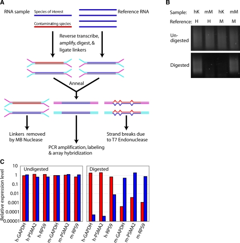
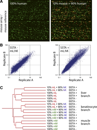
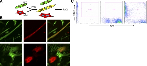
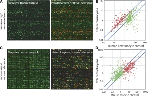
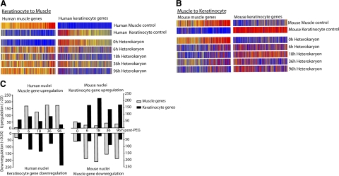
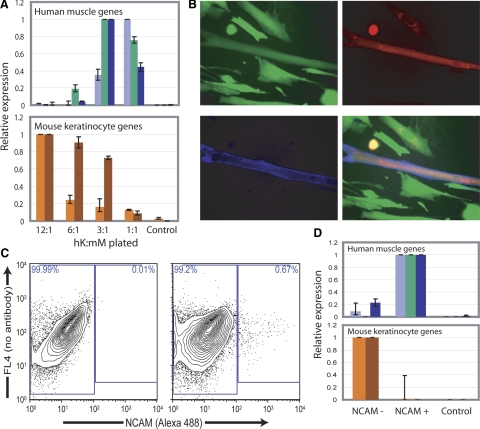
An understanding of nuclear reprogramming is fundamental to the use of cells in regenerative medicine. Due to technological obstacles, the time course and extent of reprogramming of cells following fusion has not been assessed to date. Here, we show that hundreds of genes are activated or repressed within hours of fusion of human keratinocytes and mouse muscle cells in heterokaryons, and extensive changes are observed within 4 days. This study was made possible by the development of a broadly applicable approach, species-specific transcriptome amplification (SSTA), which enables global resolution of transcripts derived from the nuclei of two species, even when the proportions of species-specific transcripts are highly skewed. Remarkably, either phenotype can be dominant; an excess of primary keratinocytes leads to activation of the keratinocyte program in muscle cells and the converse is true when muscle cells are in excess. We conclude that nuclear reprogramming in heterokaryons is rapid, extensive, bidirectional, and dictated by the balance of regulators contributed by the cell types.


Figures

Figure 1.
Figure 1.
SSTA selectively amplifies sequences from the species of interest. A) SSTA procedure scheme. RNA sample containing the species of interest (blue) and the contaminating species (red) is first converted into double-stranded DNA and ligated to specific PCR linkers. The sample is then annealed to a double-stranded cDNA reference pool of the species of interest, followed by digestion with mung bean nuclease and T7 endonuclease 1. Only the perfectly annealed duplexes in which the sample strand is from the species of interest remain undigested and are then PCR amplified. B) Human keratinocytes (hK) or mouse myoblasts (mM) were annealed to human (H) or mouse reference (M). Samples were digested with MBN/T7 endonuclease I or left undigested, and then PCR amplified for 19 cycles (undigested) or 23 cycles (digested). C) A sample containing 50% human cDNA and 50% mouse cDNA was annealed either to human reference (red bars), mouse reference (blue bars), or human + mouse reference control. Samples were digested or left undigested, and then PCR amplified. Gene abundances were assessed by species-specific quantitative PCR. Each primer pair and treatment was normalized to the human + mouse reference control.
Figure 2.
Figure 2.
Efficacy and reproducibility of SSTA. A) Human keratinocyte cDNA control (left panel) or mouse liver cDNA diluted 1:10 with human keratinocyte cDNA (right panel) were treated with SSTA using a mouse reference, labeled with Cy5 dye (red) and hybridized to microarrays along with a Cy3 common reference (green). B) Scatter plots of replicates of mouse liver (mL) cDNA diluted 1:10 with human keratinocyte (hK) cDNA either SSTA-treated (SSTA+) or untreated (SSTA−). C) Hierarchical clustering of mouse liver or mouse muscle (mM) cDNA diluted 1:10 with human cDNA or left undiluted. Replicates were hybridized to arrays with or without prior SSTA treatment. Hierarchical clustering of samples was performed using average linkage clustering.
Figure 3.
Figure 3.
Generation and isolation of heterokaryons. A) Scheme of heterokaryon formation. Human keratinocytes (dsRed) were fused with mouse myotubes (GFP) in differentiation medium, using polyethylene glycol (PEG). B) Images of GFP (green) and dsRed (red) heterokaryon cultures 4 days after fusion. Top panels: characteristic heterokaryon with myotube morphology. Bottom panels: heterokaryons with keratinocyte morphology. C) FACS profiles of human keratinocytes alone (left panel), mouse muscle cells alone (center panel), or heterokaryons (right panel). Sorting gate used for heterokaryon isolation is indicated.
Figure 4.
Figure 4.
Global analysis of reprogramming in heterokaryons. A) Mouse muscle cDNA control (left panel) or cDNA from 96 h heterokaryons (right panel) were treated with SSTA using a human reference, labeled with Cy5 dye (red) and hybridized to human microarrays along with a Cy3 common reference (green). B) Cy5/Cy3 expression values of muscle genes (red) or keratinocyte genes (green) are plotted in 4 day heterokaryons vs. human keratinocyte control. C) Human keratinocyte cDNA control (left panel) or cDNA from 96 h heterokaryons (described in A, right panel) were treated with SSTA using a mouse reference, labeled with Cy5 dye (red) and hybridized to mouse microarrays along with a Cy3 common reference (green). D) Cy5/Cy3 expression values of muscle genes (red) or keratinocyte genes (green) are plotted in 4 day heterokaryons vs. mouse muscle control.
Figure 5.
Figure 5.
Time course and extent of bidirectional reprogramming. A) Expression of muscle- or keratinocyte-specific human genes from heterokaryon profiles at various time points during reprogramming. Each vertical bar represents a gene. Red indicates high expression, blue indicates low expression. B) Expression of mouse genes in mouse muscle and keratinocyte control samples, and at various time points in heterokaryons after PEG fusion. C) Left panel: number of human muscle genes that are upregulated (≥2×) or human keratinocytes that are downregulated (≤0.5×) in comparison to human keratinocyte. Right panel: numbers of upregulated mouse keratinocytes genes and mouse muscle genes downregulated in comparison to control mouse muscle controls.
Figure 6.
Figure 6.
Reprogramming toward muscle or keratinocyte gene expression occurs in separable populations. A) Gene expression level of human muscle genes and mouse keratinocyte genes in different populations of heterokaryons produced by varying ratios of human keratinocytes to mouse myoblasts (hK:mM). Top panel: human muscle creatine kinase (light blue), myosin heavy chain 2 (green), and MyoD (dark blue), measured by quantitative RT-PCR. Bottom panel: mouse keratin 5 (orange) and periplakin (red) measured by quantitative RT-PCR. B) Expression of human muscle-specific NCAM assayed by immunohistochemistry using a human specific antibody in heterokaryons. NCAM (blue), GFP (green), dsRed (red). C) Right panel: FACS isolation of heterokaryons using a human muscle specific NCAM antibody. Left panel: mouse muscle cells are shown as a negative control. D) Reprogramming toward a muscle or keratinocyte fate assessed in NCAM+ and NCAM populations by quantitative RT-PCR. Genes assayed and controls are as in A.

References

    1. Tada M, Takahama Y, Abe K, Nakatsuji N, Tada T. Nuclear reprogramming of somatic cells by in vitro hybridization with ES cells. Curr Biol. 2001;11:1553–1558. - PubMed
    1. Cowan C A, Atienza J, Melton D A, Eggan K. Nuclear reprogramming of somatic cells after fusion with human embryonic stem cells. Science. 2005;309:1369–1373. - PubMed
    1. Silva J, Chambers I, Pollard S, Smith A. Nanog promotes transfer of pluripotency after cell fusion. Nature. 2006;441:997–1001. - PubMed
    1. Takahashi K, Yamanaka S. Induction of pluripotent stem cells from mouse embryonic and adult fibroblast cultures by defined factors. Cell. 2006;126:663–676. - PubMed
    1. Okita K, Ichisaka T, Yamanaka S. Generation of germline-competent induced pluripotent stem cells. Nature. 2007;448:313–317. - PubMed

Publication types

LinkOut - more resources