. 2009 Apr;85(4):617-26.
doi: 10.1189/jlb.1008588. Epub 2009 Jan 13.

Macrophage fusion, giant cell formation, and the foreign body response require matrix metalloproteinase 9

Affiliations

Macrophage fusion, giant cell formation, and the foreign body response require matrix metalloproteinase 9

Susan MacLauchlan et al. J Leukoc Biol. 2009 Apr.

Abstract

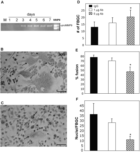
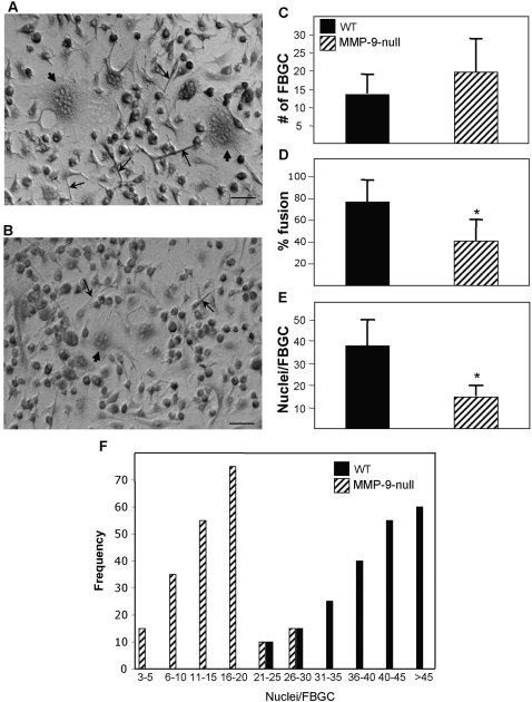
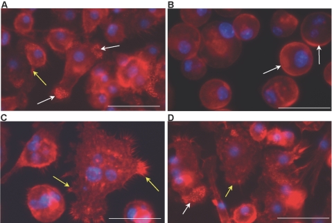
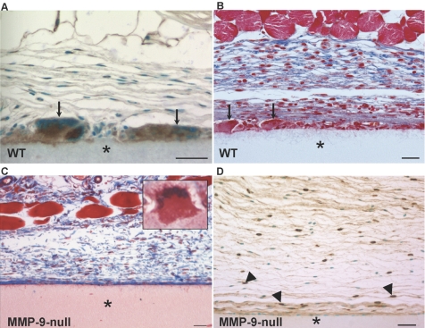
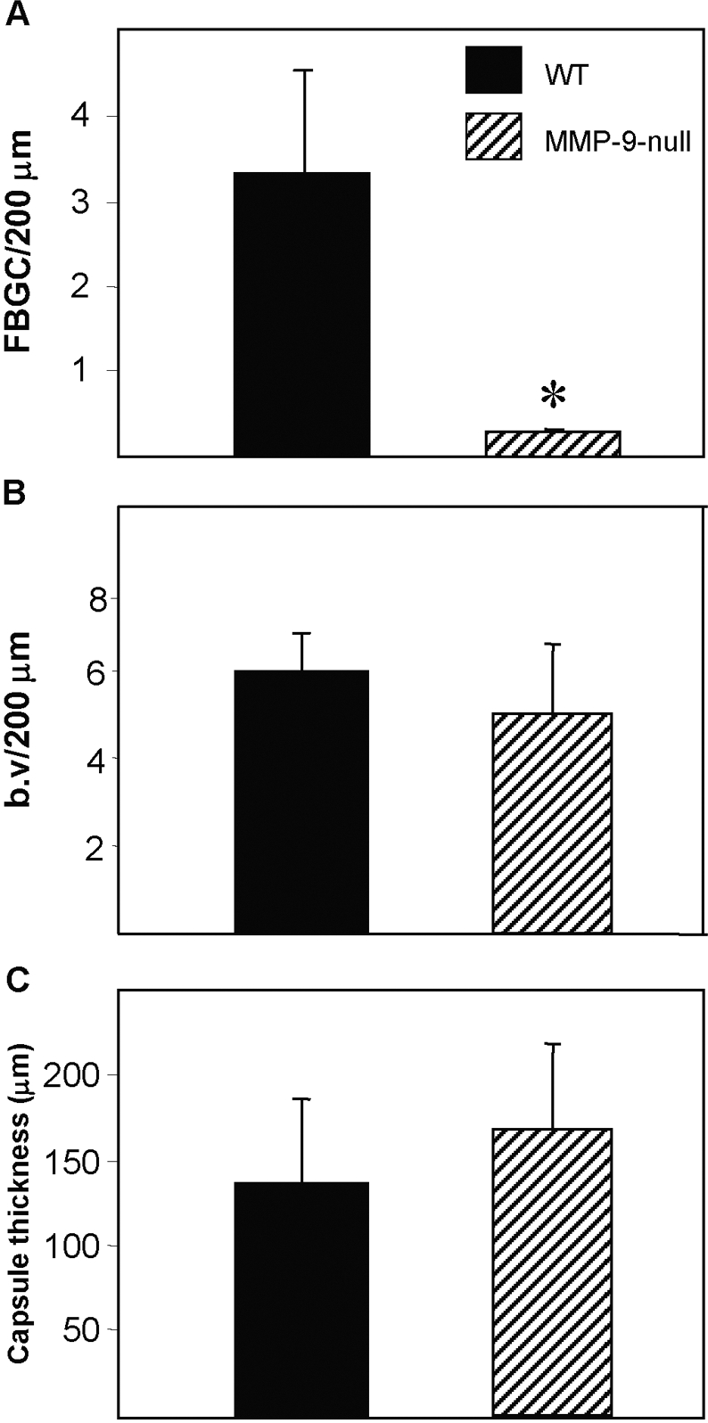
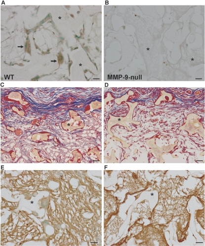
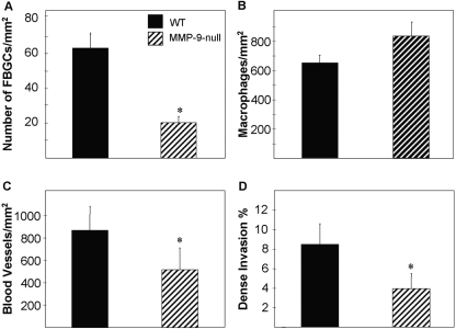
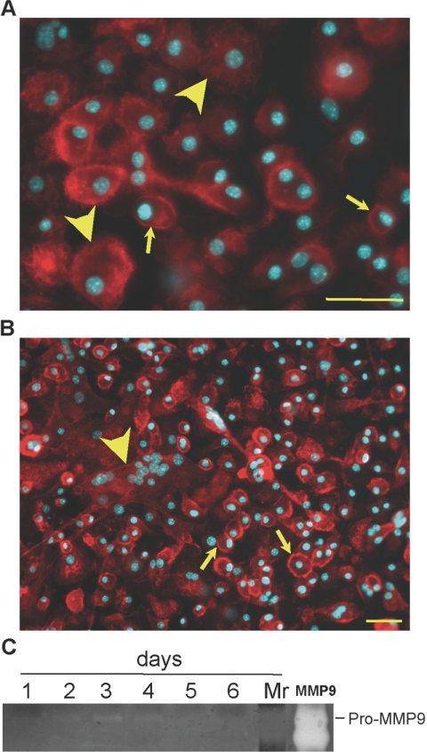
Macrophages undergo fusion to form multinucleated giant cells in several pathologic conditions, including the foreign body response (FBR). We detected high levels of matrix metalloproteinase (MMP)-9 during macrophage fusion in vitro and in foreign body giant cells (FBGCs) in vivo. Wild-type (WT) bone marrow-derived macrophages were induced to fuse with IL-4 in the presence of MMP-9 function-blocking antibodies and displayed reduced fusion. A similar defect, characterized by delayed shape change and abnormal morphology, was observed in MMP-9 null macrophages. Analysis of the FBR in MMP-9 null mice was then pursued to evaluate the significance of these findings. Specifically, mixed cellulose ester disks and polyvinyl alcohol sponges were implanted s.c. in MMP-9 null and WT mice and excised 2-4 weeks later. Histochemical and immunohistochemical analyses indicated equal macrophage recruitment between MMP-9 null and WT mice, but FBGC formation was compromised in the former. In addition, MMP-9 null mice displayed abnormalities in extracellular matrix assembly and angiogenesis. Consistent with a requirement for MMP-9 in fusion, we also observed reduced MMP-9 levels in MCP-1 null macrophages, previously shown to be defective in FBGC formation. Collectively, our studies show abnormalities in MMP-9 null mice during the FBR and suggest a role for MMP-9 in macrophage fusion.


Figures

Fig. 1.
Fig. 1.
MMP-9 expression and participation in macrophage fusion. Macrophages were stimulated to fuse with IL-4/GM-CSF, and the levels of MMP-9 were monitored by zymography. (A) Serum used in the assay was depleted of MMP-9 as shown (Lane M), and macrophages did not express MMP-9 prior to fusion (Day 1). MMP-9 was strongly induced at Day 3, and the levels remained high throughout the fusion assay. rmMMP-9 was used as positive control (Lane MMP9). (B and C). Antibody-mediated inhibition of MMP-9 reduces fusion. Representative phase-contrast images of WT macrophages induced to fuse in the presence of a function-blocking antibody (C) or isotype control IgG (B) for 7 days. Cultures treated with control IgG displayed robust FBGC formation (arrows denote FBGCs). Cultures treated with 5 μg-blocking antibody displayed formation of small FBGCs (arrows in C). Original bars = 50 μm. (D–F) Analysis of fusion at Day 7 by enumeration of various parameters in May-Grunwald-stained cultures revealed a fusion defect in antibody-treated macrophages that involved the formation of smaller FBGCs. *, P ≤ 0.05; n = 9 (D–F).
Fig. 2.
Fig. 2.
MMP-9 null macrophages display reduced fusion. Representative phase-contrast images of WT (A) and MMP-9 null macrophages (B) at Day 3 showing reduced elongation and FBGC formation in the latter. (A and B) Arrows and arrowheads denote elongated cells and FBGC, respectively. Original bars = 25 μm. (C–E) Analysis of fusion at Day 7 by enumeration of various parameters in May-Grunwald-stained cultures revealed a fusion defect in MMP-9 null macrophages that involved the formation of smaller FBGCs. *, P ≤ 0.05; n = 9. (F) Histogram demonstrating the distribution of nuclei per FBGC.
Fig. 3.
Fig. 3.
MMP-9 null macrophages display delayed and abnormal fusion-induced cytoskeletal reorganization. Representative images of WT (A and C) and MMP-9 null (B and D) IL-4-treated macrophages from Day 1 (A and B) and Day 3 (C and D) culture stained with phalloidin and DAPI are shown. At Day 1, WT macrophages exhibited normal elongation, accumulation of punctate actin (white arrows), and lamellipodia formation (yellow arrows), which were absent in MMP-9 null macrophages. Arrows in B identify the presence of peripheral actin. At Day 3, WT macrophages displayed FBGC formation and extensive lamellipodia formation (yellow arrows in C). (D) At this time-point, MMP-9 null macrophages displayed changes consistent with preparation for fusion, such as lamellipodia formation (yellow arrow) and cytoskeletal reorganization (white arrow), but FBGC formation was not evident. Original bars = 25 μm (A–D).
Fig. 4.
Fig. 4.
MMP-9 participates in FBGC formation and the FBR in vivo. (A) A section of the Millipore filter (*) implanted s.c. in WT mice for 4 weeks was stained with anti-MMP-9 antibody and visualized with the peroxidase reaction (brown color). Arrows indicate immunoreactive FBGC. (B and C) Sections of filters (*) from WT (B) and MMP-9 null (C) mice stained with Masson’s trichrome. Arrows in B denote FBGCs, which are absent in the image from MMP-9 null mice (C). (C, inset) An abnormal MMP-9 null FBGC. Examination of collagen deposition (blue color in B and C) reveals irregular assembly in MMP-9 null mice. (D) Immunohistochemical detection of macrophages with anti-Mac-3 revealed normal levels of individual macrophages (arrowheads) in MMP-9 null mice. Sections were counterstained with methyl green (A and D). Original bars = 25 μm (A–D).
Fig. 5.
Fig. 5.
MMP-9 null mice display reduced FBGC formation. Filters were implanted s.c. for 4 weeks, explanted, and analyzed by histochemistry and immunohistochemistry. (A) The number of FBGC/unit length (surface of filter) was determined and found to be significantly lower in MMP-9 null mice. *, P ≤ 0.05; n = 5. Enumeration of blood vessels (b.v; B) and capsule thickness (C) indicated normal levels in MMP-9 null mice. A total of 50 sections from five mice/group was analyzed.
Fig. 6.
Fig. 6.
Abnormal sponge granuloma formation in MMP-9 null mice. Representative images of PVA sponges (*) implanted s.c. for 2 weeks in WT (A, C, and E) and MMP-9 null (B, D, and F) mice are shown. (A) Sections stained anti-MMP-9 antibody and visualized with the peroxidase reaction (brown color) revealed high levels of expression in FBGC (arrows). (B) Background immunoreactivity was observed in MMP-9 null sections (negative control). Sections of sponges (*) from WT (C) and MMP-9 null (D) mice stained with Masson’s trichrome are shown. Examination of collagen deposition (blue color in C and D) revealed reduced deposition in MMP-9 null mice. (E and F) Immunohistochemical detection of fibronectin deposition in WT (E) and MMP-9 null (F) mice revealed abnormal deposition in the latter, characterized by thinner fibers. Sections were counterstained with methyl green (A, B, E, and F). Original bars = 25 μm (A–F).
Fig. 7.
Fig. 7.
MMP-9 null mice display reduced FBGC formation and angiogenesis in sponge granulomas. PVA sponges were implanted s.c. for 2 weeks, explanted, and analyzed by histochemistry and immunohistochemistry. (A and B) The number of FBGCs (A) and macrophages (B) per hpf was measured in Mac-3-stained sections, and the former was found to be lower in MMP-9 null mice. (C) Enumeration of blood vessels from PECAM-1-stained sections indicated a decrease in MMP-9 null mice. (D) Less-dense invasion within sponges, determined from Masson’s trichrome-stained sections, was observed in MMP-9 null mice. A total of 50 sections from five mice/group was analyzed. *, P ≤ 0.05; n = 5.
Fig. 8.
Fig. 8.
Reduced MMP-9 levels in fusion-defective MCP-1 null macrophages. Representative images of MCP-1 null macrophages induced to fuse with IL-4 for 2 (A) and 7 days (B) and visualized with rhodamine-phalloidin and DAPI. (A) Arrows and arrowheads denote single cells with peripheral and cytoplasmic actin distribution, respectively. (B) Formation of FBGC (arrowhead) and single cells with peripheral actin (arrows) can be observed in Day 7 cultures. Original bars = 50 μm. (C) The levels of MMP-9 were monitored by zymography and revealed that MCP-1 null macrophages express low levels of MMP-9 during fusion. rmMMP-9 (lane MMP9) was used as positive control. Lane Mr, molecular weight standards.

References

    1. Sapir A, Avinoam O, Podbilewicz B, Chernomordik L V. Viral and developmental cell fusion mechanisms: conservation and divergence. Dev Cell. 2008;14:11–21. - PMC - PubMed
    1. Oren-Suissa M, Podbilewicz B. Cell fusion during development. Trends Cell Biol. 2007;17:537–546. - PubMed
    1. Chen E H, Olson E N. Unveiling the mechanisms of cell-cell fusion. Science. 2005;308:369–373. - PubMed
    1. Vignery A. Macrophage fusion: the making of osteoclasts and giant cells. J Exp Med. 2005;202:337–340. - PMC - PubMed
    1. Anderson J M, Rodriguez A, Chang D T. Foreign body reaction to biomaterials. Semin Immunol. 2008;20:86–100. - PMC - PubMed

Publication types