. 2009 Jan;21(1):334-46.
doi: 10.1105/tpc.108.064360. Epub 2009 Jan 13.

STARCH-EXCESS4 is a laforin-like Phosphoglucan phosphatase required for starch degradation in Arabidopsis thaliana

Affiliations

STARCH-EXCESS4 is a laforin-like Phosphoglucan phosphatase required for starch degradation in Arabidopsis thaliana

Oliver Kötting et al. Plant Cell. 2009 Jan.

Abstract

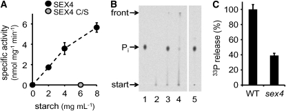
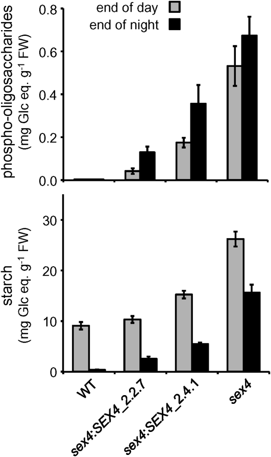
Starch is the major storage carbohydrate in plants. It is comprised of glucans that form semicrystalline granules. Glucan phosphorylation is a prerequisite for normal starch breakdown, but phosphoglucan metabolism is not understood. A putative protein phosphatase encoded at the Starch Excess 4 (SEX4) locus of Arabidopsis thaliana was recently shown to be required for normal starch breakdown. Here, we show that SEX4 is a phosphoglucan phosphatase in vivo and define its role within the starch degradation pathway. SEX4 dephosphorylates both the starch granule surface and soluble phosphoglucans in vitro, and sex4 null mutants accumulate phosphorylated intermediates of starch breakdown. These compounds are linear alpha-1,4-glucans esterified with one or two phosphate groups. They are released from starch granules by the glucan hydrolases alpha-amylase and isoamylase. In vitro experiments show that the rate of starch granule degradation is increased upon simultaneous phosphorylation and dephosphorylation of starch. We propose that glucan phosphorylating enzymes and phosphoglucan phosphatases work in synergy with glucan hydrolases to mediate efficient starch catabolism.


Figures

Figure 1.
Figure 1.
SEX4 Dephosphorylates the Starch Granule Surface. (A) Specific activities of recombinant SEX4 (black circles; 0.5 μg mL−1) and the active site mutant SEX4 C/S (gray circle; 0.5 μg mL−1) incubated with different amounts of native starch granules that had been prephosphorylated in vitro by recombinant GWD and [β-33P]ATP as phosphate donor. Activity was measured as the release of 33P from the granules into the supernatant. Values are means ± se; n = 3 independent experiments. (B) Thin layer chromatography of the products released from 33P-labeled starch granules by recombinant SEX4 protein (as in [A]) and leaf extracts of wild-type and sex4 plants (as in [C]). Two microliters of each were analyzed as described (Hejazi et al., 2008). Arrows indicate start, front, and the position of orthophosphate. 1, recombinant SEX4; 2, medium control; 3, wild-type extract; 4, sex4 extract; 5, hydrolyzed [β-33P]ATP. All samples were analyzed on the same plate. (C) Quantification of released 33P from in vitro prephosphorylated starch granules (as in [A]) by crude extracts from leaves of wild-type and sex4 plants (0.22 mg total protein mL−1). Values are means ± se; n = 5 independent experiments.
Figure 2.
Figure 2.
Phospho-Oligosaccharides Accumulate in Mutants Lacking SEX4. (A) HPAEC-PAD analysis of wild-type and sex4 extracts. Leaves were harvested at the end of the night and extracted in perchloric acid. Prior to purification by ion-exchange chromatography, neutralized extracts were treated with (+) or without (−) Antarctic Phosphatase and subsequently subjected to HPAEC-PAD analysis. One representative chromatogram each is shown (n = 5 individual plants). Numbers above the panel indicate the DP of the detected glucan chains. Essentially the same result was obtained when performing the phosphatase treatment with recombinant SEX4 instead of Antarctic Phosphatase (see Supplemental Figure 1 online). (B) MS analysis of phospho-oligosaccharides enriched from sex4 extracts. Whole rosettes of sex4 plants were extracted in hot ethanol. Phospho-oligosaccharides were enriched on a Carbograph column and subjected to MS analysis. The mass-to-charge (m/z) values obtained reflect the theoretical masses of phospho-oligosaccharides with a DP of 5 to 9 glucose units, comprising one (1P) or two (2P) phosphate groups. Analytes were recovered as monosodium (Na+) or monoprotonated (H+) adduct ions. No phospho-oligosaccharides could be detected in wild-type extracts.
Figure 3.
Figure 3.
Phospho-Oligosaccharides in sex4 Are Produced at Night and Accumulate during Development. (A) Phospho-oligosaccharides (open symbols, right scale) and starch (closed symbols, left scale) levels of sex4 (triangles) and wild-type (squares) plants were determined at different times during the diurnal cycle. Values are means ± se; n = 5 individual plants. Error bars not visible are smaller than the symbols. Values for 0 h are replotted from the 24 h time point and so do not represent actual values; indicated by the dashed lines. FW, fresh weight. (B) Whole rosettes comprising 18 to 24 leaves of wild-type and sex4 plants were harvested at the end of the day and divided into two fractions. The first fraction comprised the eight youngest visible leaves pooled from two individual plants (young), and the second fraction contained the rest of the leaves from one plant (old). Starch (black bars; left scale) and phospho-oligosaccharide (gray bars; right scale) contents were measured. Values are means ± se; n = 5 biological replicates.
Figure 4.
Figure 4.
Expression of the SEX4 cDNA Partially Complements the sex4 Phenotype. Phospho-oligosaccharides and starch contents at the end of the day (gray bars) and the end of the night (black bars) were determined in wild-type and sex4 plants as well as in two independent sex4 lines expressing the SEX4 cDNA under control of the cauliflower mosaic virus 35S promoter (sex4:SEX4_2.2.7, sex4:SEX4_2.4.1). Values represent the mean ± se of at least five biological replicates.
Figure 5.
Figure 5.
Hydrolytic Glucan Release Is Stimulated upon Simultaneous Phosphorylation and Dephosphorylation of Starch. Phosphate-free starch granules from GWD-deficient sex1-3 mutants were incubated with recombinant enzymes in the presence of 1 mM ATP. Glucan release into the supernatant was determined after acid hydrolysis. Since the absolute values varied significantly between individual experiments, glucan release was normalized to the maximum glucan release. Values are means ± se; n = 3 independent experiments, each with triplicate measurements.
Figure 6.
Figure 6.
Mutation of AMY3, ISA3, PGM, or GWD Alters the Phenotype of sex4 Plants. Phospho-oligosaccharide and starch contents of 4-week-old plants (wild type and indicated mutants) were determined at the end of the day (gray bars) and the end of the night (black bars). Values are means ± se; n = 5 individual plants.
Figure 7.
Figure 7.
Phospho-Oligosaccharide Pattern in sex4 Plants Is Altered upon Loss of AMY3 or ISA3 Expression. Representative HPAEC-PAD chromatograms (n = 5 individual plants) of phosphatase-treated extracts from wild-type, sex4, amy3, isa3, and the respective double mutant plants harvested at the end of the night. Numbers above the panel indicate the DP of the detected (phospho-) glucans. The panel on the right represents a zoom into the region of late-eluting glucans.
Figure 8.
Figure 8.
Proposed Model of the Initial Events at the Granule Surface during Starch Breakdown. At night, starch is phosphorylated at the granule surface by GWD and PWD in both wild-type and sex4 plants (A), leading to partial unwinding of amylopectin double helices. In the wild type, BAM3 and SEX4 can subsequently release maltose and phosphate, respectively (B), while ISA3 hydrolyzes branch points and releases malto-oligosaccharides (C). In sex4 mutants, phosphate is not removed by SEX4, leading to reduced maltose release by BAM3 (D). The following actions of ISA3 and AMY3 release malto- and phospho-oligosaccharides. After degradation of one semicrystalline lamella, a new cycle can start again with the phosphorylation of the granule surface by GWD and PWD (A).

References

    1. Alonso, A., Sasin, J., Bottini, N., Friedberg, I., Friedberg, I., Osterman, A., Godzik, A., Hunter, T., Dixon, J., and Mustelin, T. (2004). Protein tyrosine phosphatases in the human genome. Cell 117 699–711. - PubMed
    1. Baunsgaard, L., Lütken, H., Mikkelsen, R., Glaring, M.A., Pham, T.T., and Blennow, A. (2005). A novel isoform of glucan, water dikinase phosphorylates pre-phosphorylated α-glucans and is involved in starch degradation in Arabidopsis. Plant J. 41 595–605. - PubMed
    1. Blennow, A., Nielsen, T.H., Baunsgaard, L., Mikkelsen, R., and Engelsen, S.B. (2002). Starch phosphorylation: A new front line in starch research. Trends Plant Sci. 7 445–450. - PubMed
    1. Caspar, T., Huber, S.C., and Somerville, C. (1985). Alterations in growth, photosynthesis, and respiration in a starchless mutant of Arabidopsis thaliana (L.) deficient in chloroplast phosphoglucomutase activity. Plant Physiol. 79 11–17. - PMC - PubMed
    1. Delatte, T., Umhang, M., Trevisan, M., Eicke, S., Thorneycroft, D., Smith, S.M., and Zeeman, S.C. (2006). Evidence for distinct mechanisms of starch granule breakdown in plants. J. Biol. Chem. 281 12050–12059. - PubMed

Publication types

MeSH terms