Comparative Study
. 2009 Apr;329(1):2-13.
doi: 10.1124/jpet.108.148270. Epub 2009 Jan 13.

Characterization of the A2B adenosine receptor from mouse, rabbit, and dog

Affiliations
Comparative Study

Characterization of the A2B adenosine receptor from mouse, rabbit, and dog

John A Auchampach et al. J Pharmacol Exp Ther. 2009 Apr.

Abstract

We have cloned and pharmacologically characterized the A(2B) adenosine receptor (AR) from the dog, rabbit, and mouse. The full coding regions of the dog and mouse A(2B)AR were obtained by reverse transcriptase-polymerase chain reaction, and the rabbit A(2B)AR cDNA was obtained by screening a rabbit brain cDNA library. It is noteworthy that an additional clone was isolated by library screening that was identical in sequence to the full-length rabbit A(2B)AR, with the exception of a 27-base pair deletion in the region encoding amino acids 103 to 111 (A(2B)AR(103-111)). This 9 amino acid deletion is located in the second intracellular loop at the only known splice junction of the A(2B)AR and seems to result from the use of an additional 5' donor site found in the rabbit and dog but not in the human, rat, or mouse sequences. [(3)H]3-Isobutyl-8-pyrrolidinoxanthine and 8-[4-[((4-cyano-[2,6-(3)H]-phenyl)carbamoylmethyl)oxy]phenyl]-1,3-di(n-propyl)xanthine ([(3)H]MRS 1754) bound with high affinity to membranes prepared from human embryonic kidney (HEK) 293 cells expressing mouse, rabbit, and dog A(2B)ARs. Competition binding studies performed with a panel of agonist (adenosine and 2-amino-3,5-dicyano-4-phenylpyridine analogs) and antagonist ligands identified similar potency orders for the A(2B)AR orthologs, although most xanthine antagonists displayed lower binding affinity for the dog A(2B)AR compared with A(2B)ARs from rabbit and mouse. No specific binding could be detected with membranes prepared from HEK 293 cells expressing the rabbit A(2B)AR(103-111) variant. Furthermore, the variant failed to stimulate adenylyl cyclase or calcium mobilization. We conclude that significant differences in antagonist pharmacology of the A(2B)AR exist between species and that some species express nonfunctional variants of the A(2B)AR due to "leaky" splicing.


Figures

Fig. 1.
Fig. 1.
Synthetic routes to 6-amino-3,5-dicyano-4-phenylpyridine derivatives. Benzaldehyde or 4-hydroxybenzaldehyde was reacted with cyanothioacetamide and a catalytic amount of triethylamine (Et3-N) in ethanol to generate compounds 1 and 2. Compound 2 was further reacted with 2-chloroacetamide and sodium hydrogen carbonate in dimethyl formamide (DMF) to generate compound 3. The compounds were decolorized with activated carbon and recrystallized from methanol or ethanol. Identity of the compounds was performed by melting point analysis, infrared spectroscopy, and NMR. Compound 4 (BAY 60-6583) was provided by Bayer Healthcare.
Fig. 2.
Fig. 2.
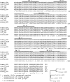
Amino acid alignment of the human, rabbit, dog, rat, and mouse A2BAR sequences. Solids lines indicate putative transmembrane domains (TM) with numbered designations (I-VII). This figure was prepared using Biology Workbench 3.2 (San Diego Supercomputing Center).
Fig. 3.
Fig. 3.
A, schematic illustration of the genomic organization of the A2BAR depicting the scheme for formation of the A2BAR103-111 variant during splicing by use of an alternative 5′ donor site. B, nucleotide sequence alignment of the second intracellular loop of the mouse, rat, human, dog, and rabbit A2BAR sequences as well as the A2BAR103-111 variant. Proximal sequences of the 5′ and 3′ ends of the introns (lowercase letters) for the mouse (GenBank accession number NC_000077), rat (NC_005109), and human A2BARs (NC_000017) are shown. The traditional 5′ and 3′ donor sites are shown with the exonic and intronic portions depicted in blue and red, respectively. The alternative 5′ donor site found in the rabbit and dog sequences is also shown. C, amino acid alignment of the second intracellular loop of the various species showing the 9 amino acid deletion in the rabbit A2BAR103-111 variant. The conserved DRY sequence is highlighted in red.
Fig. 4.
Fig. 4.
Trace detection of the A2BAR103-111 variant in rabbit brain by RT-PCR. Total RNA obtained from rabbit (top gel) or mouse (bottom gel) brain was reverse-transcribed, subjected to 40 cycles of PCR amplification using species-specific primers spanning the splicing region, and subjected to gel electrophoresis, as described under Materials and Methods. The lane labeled Control contained reactions that included full-length (mouse) or a mixture of full-length and A2BAR103-111 variant cDNAs (rabbit), and the lane labeled H2O contained reactions using water in place of cDNA. For studies using rabbit cDNA, a 171-bp fragment corresponds to the full-length receptor, and a 144-bp fragment corresponds to the splice variant. Only a single 158-bp fragment was detected in studies using mouse cDNA, which corresponds to the full-length mouse A2BAR receptor.
Fig. 5.
Fig. 5.
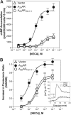
Changes in intracellular levels of cAMP (A) and Ca2+ (B) in HEK 293 cells transfected with the empty vector (pcDNA3.1), the full-length rabbit A2BAR, or the rabbit A2BAR103-111 variant in response to increasing concentrations of NECA. For cAMP assays, EC50 (nM; n = 6) and Emax (pmol/50,000 cells/15 min) values were: vector = 2050 ± 1020 and 2145 ± 364; A2BAR = 108 ± 41 and 6932 ± 508; and A2BAR103-111 = 2931 ± 940 and 1814 ± 218. For Ca2+ assays, EC50 (nM; mean ± S.E.M.; n = 6) and Emax (peak increase in fluorescence ratio from baseline) values were: vector = 1024 ± 440 and 0.69 ± 0.09; A2BAR = 125 ± 38 and 1.00 ± 0.05; and A2BAR103-111, 779 ± 292 and 0.73 ± 0.08. B, inset, the effect of 1 μM NECA on intracellular [Ca2+] calculated from the fluorescence ratio in the three lines of transfected HEK 293 cells. Note that baseline intracellular Ca2+ levels are increased in HEK 293 cells transfected with the full-length A2BAR compared with cells transfected with either the empty vector or the A2BAR103-111 variant.
Fig. 6.
Fig. 6.
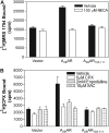
One-point radioligand binding assays using cell membranes obtained from HEK 293 cells transfected with the empty vector (pcDNA3.1), the full-length rabbit A2BAR, or the rabbit A2BAR103-111 variant. Significant specific binding with [3H]MRS 1754 (A) and [3H]CPX (B) was observed in assays using cell membranes obtained from cells transfected with the full-length rabbit A2BAR, but not with the A2BAR103-111 variant, using 100 μM NECA, 5 μM CPX, 2 mM enprofylline, or 10 μM XAC to define nonspecific binding. The reactions (3-h incubations with 100-μg membrane protein/tube) included 6 nM [3H]MRS 1754 or 30 nM [3H]CPX. Data are means ± S.E.M. of triplicate determinations. The results are representative of three experiments.
Fig. 7.
Fig. 7.
Confocal images of HEK 293 cell lines expressing the full-length rabbit A2BAR or the rabbit A2BAR103-111 variant fused at the C terminus with GFP.
Fig. 8.
Fig. 8.
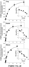
Saturation isotherm of [3H]MRS 1754 binding to dog, rabbit, and mouse A2BARs expressed in HEK 293 cells. Insets depict Scatchard transformation of the data. [3H]MRS 1754 (0.5-20 nM) was incubated with 50-μg membrane protein for 3 h at room temperature. Specific binding data, obtained by subtracting nonspecific binding defined by inclusion of 100 μM NECA, is shown. Values are means ± S.E.M. of triplicate determinations. The results are representative of three experiments.
Fig. 9.
Fig. 9.
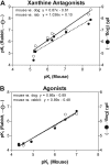
Plot of affinities of xanthine antagonists (A) and AR agonists (B) for the mouse A2BAR versus the rabbit and dog A2BARs. The results of regression analysis are reported within the graphs.
Fig. 10.
Fig. 10.
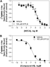
Competition for [3H]MRS 1754 binding to membranes from HEK 293 cells expressing the mouse A2BAR by NECA (A) or compound 3 (B) in the presence of vehicle or 100 μM guanosine 5′-O-(γ-thio)triphosphate (GTPγS). The reactions (3-h incubations with 100-μg membrane protein/tube) included 6 nM [3H]MRS 1754. Specific binding was defined by use of 100 μM NECA. Data (n = 5) are means ± S.E.M.

References

    1. Auchampach JA, Jin X, Wan TC, Caughey GH, and Linden J (1997) Canine mast cell adenosine receptors: cloning and expression of the A3 receptor and evidence that degranulation is mediated by the A2B receptor. Mol Pharmacol 52 846-860. - PubMed
    1. Baraldi PG, Tabrizi MA, Fruttarolo F, Romagnoli R, and Preti D (2008) Recent improvements in the development of A2B adenosine receptor agonists. Purinergic Signal 4 287-303. - PMC - PubMed
    1. Beukers MW, Chang LC, von Frijtag Drabbe Künzel JK, Mulder-Krieger T, Spanjersberg RF, Brussee J, and IJzerman AP (2004) New, non-adenosine, high-potency agonists for the human adenosine A2B receptor with an improved selectivity profile compared to the reference agonist N-ethylcarboxamidoadenosine. J Med Chem 47 3707-3709. - PubMed
    1. Cherezov V, Rosenbaum DM, Hanson MA, Rasmussen SG, Thian FS, Kobilka TS, Choi HJ, Kuhn P, Weis WI, Kobilka BK, et al. (2007) High-resolution crystal structure of an engineered human β2-adrenergic G protein-coupled receptor. Science 318 1258-1265. - PMC - PubMed
    1. Cooper J, Hill SJ, and Alexander SP (1997) An endogenous A2B adenosine receptor coupled to cyclic AMP generation in human embryonic kidney (HEK 293) cells. Br J Pharmacol 122 546-550. - PMC - PubMed

Publication types

MeSH terms

LinkOut - more resources