A mutation in the mouse Chd2 chromatin remodeling enzyme results in a complex renal phenotype
- PMID: 19142019
- PMCID: PMC2818461
- DOI: 10.1159/000190788
A mutation in the mouse Chd2 chromatin remodeling enzyme results in a complex renal phenotype
Abstract
Background and aims: Glomerular diseases are the third leading cause of kidney failure worldwide, behind only diabetes and hypertension. The molecular mechanisms underlying the cause of glomerular diseases are still largely unknown. The identification and characterization of new molecules associated with glomerular function should provide new insights into understanding the diverse group of glomerular diseases. The Chd2 protein belongs to a family of enzymes involved in ATP-dependent chromatin remodeling, suggesting that it likely functions as an epigenetic regulator of gene expression via the modification of chromatin structure.
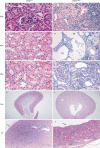
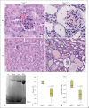
Methods: In this study, we present a detailed histomorphologic characterization of mice containing a mutation in the chromodomain helicase DNA-binding protein 2 (Chd2).
Results: We show that Chd2-mutant mice present with glomerulopathy, proteinuria, and significantly impaired kidney function. Additionally, serum analysis revealed decreased hemoglobin and hematocrit levels in Chd2-mutant mice, suggesting that the glomerulopathy observed in these mice is associated with anemia.
Conclusion: Collectively, the data suggest a role for the Chd2 protein in the maintenance of kidney function.
Copyright 2009 S. Karger AG, Basel.
Figures


Similar articles
-
Human CHD2 is a chromatin assembly ATPase regulated by its chromo- and DNA-binding domains.J Biol Chem. 2015 Jan 2;290(1):25-34. doi: 10.1074/jbc.M114.609156. Epub 2014 Nov 10. J Biol Chem. 2015. PMID: 25384982 Free PMC article.
-
Mutations in CHD2 cause defective association with active chromatin in chronic lymphocytic leukemia.Blood. 2015 Jul 9;126(2):195-202. doi: 10.1182/blood-2014-10-604959. Epub 2015 Jun 1. Blood. 2015. PMID: 26031915
-
Mutation of the SNF2 family member Chd2 affects mouse development and survival.J Cell Physiol. 2006 Oct;209(1):162-71. doi: 10.1002/jcp.20718. J Cell Physiol. 2006. PMID: 16810678
-
Chromatin Remodeling Proteins in Epilepsy: Lessons From CHD2-Associated Epilepsy.Front Mol Neurosci. 2018 Jun 15;11:208. doi: 10.3389/fnmol.2018.00208. eCollection 2018. Front Mol Neurosci. 2018. PMID: 29962935 Free PMC article. Review.
-
CHD2-Related CNS Pathologies.Int J Mol Sci. 2021 Jan 8;22(2):588. doi: 10.3390/ijms22020588. Int J Mol Sci. 2021. PMID: 33435571 Free PMC article. Review.
Cited by
-
Recent Advances in Glioma Cancer Treatment: Conventional and Epigenetic Realms.Vaccines (Basel). 2022 Sep 2;10(9):1448. doi: 10.3390/vaccines10091448. Vaccines (Basel). 2022. PMID: 36146527 Free PMC article. Review.
-
Autism-linked CHD gene expression patterns during development predict multi-organ disease phenotypes.J Anat. 2018 Dec;233(6):755-769. doi: 10.1111/joa.12889. Epub 2018 Oct 2. J Anat. 2018. PMID: 30277262 Free PMC article.
-
The role of epigenetics in the pathology of diabetic complications.Am J Physiol Renal Physiol. 2010 Jul;299(1):F14-25. doi: 10.1152/ajprenal.00200.2010. Epub 2010 May 12. Am J Physiol Renal Physiol. 2010. PMID: 20462972 Free PMC article. Review.
-
Potential Epigenetic-Based Therapeutic Targets for Glioma.Front Mol Neurosci. 2018 Nov 15;11:408. doi: 10.3389/fnmol.2018.00408. eCollection 2018. Front Mol Neurosci. 2018. PMID: 30498431 Free PMC article. Review.
-
Epigenetic DNA Methylation and Protein Homocysteinylation: Key Players in Hypertensive Renovascular Damage.Int J Mol Sci. 2024 Oct 29;25(21):11599. doi: 10.3390/ijms252111599. Int J Mol Sci. 2024. PMID: 39519150 Free PMC article. Review.
References
-
- El Nahas M. The global challenge of chronic kidney disease. Kidney Int. 2005;68:2918–2929. - PubMed
-
- US Renal Data System, USRDS 2006 Annual Data Report: Atlas of End-Stage Renal Disease in the United States. Bethesda: National Institutes of Health, National Institute of Diabetes and Digestive and Kidney Diseases; 2006.
-
- Miniño AM, Heron MP, Smith BL. Deaths: preliminary data for 2004. Natl Vital Stat Rep. 2006;54:1–49. - PubMed
-
- Lysaght MJ. Maintenance dialysis population dynamics: current trends and long-term implications. J Am Soc Nephrol. 2002;13(suppl 1):S37–S40. - PubMed
-
- Xue JL, Ma JZ, Louis TA, Collins AJ. Forecast of the number of patients with end-stage renal disease in the United States to the year 2010. J Am Soc Nephrol. 2001;12:2753–2758. - PubMed
Publication types
MeSH terms
Substances
Grants and funding
LinkOut - more resources
Full Text Sources
Medical
Molecular Biology Databases

