

Review
doi: 10.1038/nrc2462.
Optical imaging for cervical cancer detection: solutions for a continuing global problem
Affiliations
- PMID: 19143057
- PMCID: PMC2633464
- DOI: 10.1038/nrc2462
Item in Clipboard
Review
Optical imaging for cervical cancer detection: solutions for a continuing global problem
Nat Rev Cancer.
2008 Sep.
Abstract
Cervical cancer is the leading cause of cancer death for women in developing countries. Optical technologies can improve the accuracy and availability of cervical cancer screening. For example, battery-powered digital cameras can obtain multi-spectral images of the entire cervix, highlighting suspicious areas, and high-resolution optical technologies can further interrogate such areas, providing in vivo diagnosis with high sensitivity and specificity. In addition, targeted contrast agents can highlight changes in biomarkers of cervical neoplasia. Such advances should provide a much needed global approach to cervical cancer prevention.
Figures
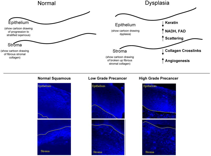
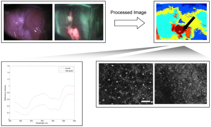
Top row: Widefield reflectance and autofluorescence imaging can interrogate the entire field of the cervix, indicating suspicious areas; digital image analysis approaches can help to objectify and automate the recognition of abnormal areas with high sensitivity. High spectral or spatial resolution techniques can be used to probe suspicious areas to confirm diagnosis of precancerous areas. Spectroscopy (lower left) can probe changes in the concentration of tissue chromophores, while confocal microscopy (lower right) can directly images changes in cell morphology and nuclear to cytoplasmic ratio without the need to biopsy, section and stain tissue. Scale bars measure 1mm in the widefield images, and 50 um in the confocal images.
References
-
- Parkin DM, Bray F, Ferlay J, Pisani P. Global cancer statistics, 2002. CA: a cancer journal for clinicians. 2005;55:74–108. - PubMed
-
- Koss LG. The Papanicolaou test for cervical cancer detection. A triumph and a tragedy. JAMA. 1989;261:737–43. - PubMed
-
- Walboomers JM, et al. Human papillomavirus is a necessary cause of invasive cervical cancer worldwide. J Pathol. 1999;189:12–9. - PubMed
-
- Cohen J. Public health. High hopes and dilemmas for a cervical cancer vaccine. Science. 2005;308:618–21. - PubMed
-
- Agosti JM, Goldie SJ. Introducing HPV vaccine in developing countries--key challenges and issues. N Engl J Med. 2007;356:1908–10. - PubMed
Publication types
MeSH terms
Substances
Grants and funding
LinkOut - more resources
Full Text Sources
Other Literature Sources
Medical
