. 2009 Jan 13;6(1):e1.
doi: 10.1371/journal.pmed.0060001.

Ectopic lymphoid structures support ongoing production of class-switched autoantibodies in rheumatoid synovium

Affiliations

Ectopic lymphoid structures support ongoing production of class-switched autoantibodies in rheumatoid synovium

Frances Humby et al. PLoS Med. .

Abstract

Background: Follicular structures resembling germinal centres (GCs) that are characterized by follicular dendritic cell (FDC) networks have long been recognized in chronically inflamed tissues in autoimmune diseases, including the synovium of rheumatoid arthritis (RA). However, it is debated whether these ectopic structures promote autoimmunity and chronic inflammation driving the production of pathogenic autoantibodies. Anti-citrullinated protein/peptide antibodies (ACPA) are highly specific markers of RA, predict a poor prognosis, and have been suggested to be pathogenic. Therefore, the main study objectives were to determine whether ectopic lymphoid structures in RA synovium: (i) express activation-induced cytidine deaminase (AID), the enzyme required for somatic hypermutation and class-switch recombination (CSR) of Ig genes; (ii) support ongoing CSR and ACPA production; and (iii) remain functional in a RA/severe combined immunodeficiency (SCID) chimera model devoid of new immune cell influx into the synovium.

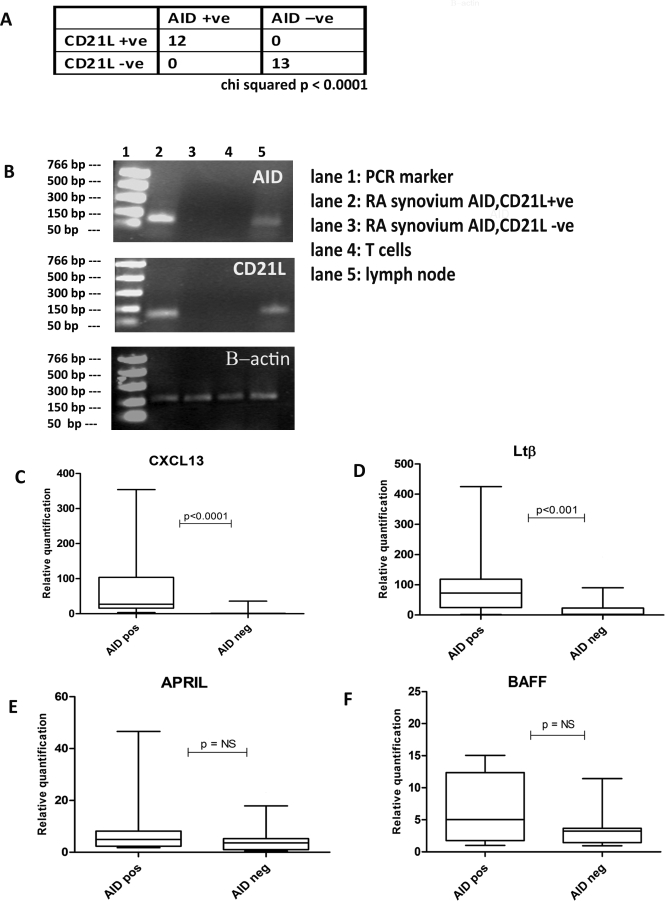
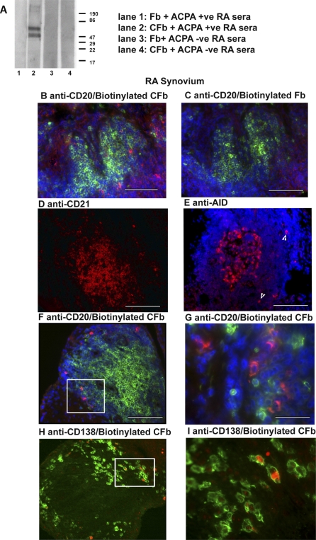
Methods and findings: Using immunohistochemistry (IHC) and quantitative Taqman real-time PCR (QT-PCR) in synovial tissue from 55 patients with RA, we demonstrated that FDC+ structures invariably expressed AID with a distribution resembling secondary lymphoid organs. Further, AID+/CD21+ follicular structures were surrounded by ACPA+/CD138+ plasma cells, as demonstrated by immune reactivity to citrullinated fibrinogen. Moreover, we identified a novel subset of synovial AID+/CD20+ B cells outside GCs resembling interfollicular large B cells. In order to gain direct functional evidence that AID+ structures support CSR and in situ manufacturing of class-switched ACPA, 34 SCID mice were transplanted with RA synovium and humanely killed at 4 wk for harvesting of transplants and sera. Persistent expression of AID and Igamma-Cmu circular transcripts (identifying ongoing IgM-IgG class-switching) was observed in synovial grafts expressing FDCs/CD21L. Furthermore, synovial mRNA levels of AID were closely associated with circulating human IgG ACPA in mouse sera. Finally, the survival and proliferation of functional B cell niches was associated with persistent overexpression of genes regulating ectopic lymphoneogenesis.

Conclusions: Our demonstration that FDC+ follicular units invariably express AID and are surrounded by ACPA-producing plasma cells provides strong evidence that ectopic lymphoid structures in the RA synovium are functional and support autoantibody production. This concept is further confirmed by evidence of sustained AID expression, B cell proliferation, ongoing CSR, and production of human IgG ACPA from GC+ synovial tissue transplanted into SCID mice, independently of new B cell influx from the systemic circulation. These data identify AID as a potential therapeutic target in RA and suggest that survival of functional synovial B cell niches may profoundly influence chronic inflammation, autoimmunity, and response to B cell-depleting therapies.


Conflict of interest statement

Competing Interests: The authors have declared that no competing interests exist.

Figures

Figure 1
Figure 1. AID Expression within the Rheumatoid Synovial Membrane Is Restricted to Lymphoid Aggregates with FDC Networks
A representative example of sequential paraffin-embedded sections of LN and RA synovial membrane stained for AID (brown, A, E, I), T cells (CD3, brown, B, F, J), B cells (CD20, red, C,G, K) and follicular DCs (CD21, red, D,H, L). The presence of FDC networks was invariably associated with the expression of AID even in FDC+ grade 3 aggregates (arrow, A), without morphologically detectable dark and light zones within the B cell rich area (A−D). In more organised grade 3 aggregates (E−H), expression was more prominent and closely resembled that seen in secondary lymphoid organs, such as the LN (I−L) (original magnification was 20×). Scale bars 200 μm.
Figure 2
Figure 2. AID mRNA within Rheumatoid Synovial Tissue Is Expressed Exclusively in Association with CD21L-Isoform Transcripts
QT-PCR was used to measure levels of transcripts of AID and CD21L in synovial samples from 25 patients with RA. Results were normalized for the endogenous control (human β-actin) and expressed as relative quantification. (A) The presence of AID is restricted to those tissues expressing CD21L. (B) Representative examples of the PCR products were run in a 1.8% agarose gel to ensure the presence of a single specific amplification product and confirm specficity of QT-PCR. Amplicons of the expected length (99 bp for AID and CD21L and 171 bp for β-actin) were observed in samples that gave positive signals at QT-PCR (lane 2), while no bands were detected in samples negative for AID/CD21L by QT-PCR (lane 3). No AID/CD21L amplification was detectable in negative controls (CD3-sorted cells, lane 4), while AID and CD21L were both detectable in control human lymph node (lane 5). (C–F) CXCL13, LTβ, BAFF, and APRIL transcript analysis by QT-PCR. AID expression within synovial tissue was associated with the up-regulation of both CXCL13 and LTβ but no significant difference was seen in the levels of APRIL and BAFF. The lower and upper margins of the box represent the 25th and 75th percentiles, with the extended arms representing the 10th and 90th percentiles, respectively. The median is shown as a horizontal line within the box. p-Values were calculated using the Mann-Whitney U test.
Figure 3
Figure 3. AID Expression within the Rheumatoid Synovial Membrane Identifies Interfollicular Large B Cells
(A) Paraffin-embedded sections from RA patients were stained for AID (20× magnification). AID+ cells with a large cytoplasm and sometimes dendritic-like morphology (arrows) were frequently and exclusively seen in RA synovial tissue characterized by the presence of large aggregates predominantly in close proximity to CD21+ FDC networks. (B) Paraffin sections were double stained for AID (brown) and CD3 (red) (60× magnification), demonstrating the close relationship between AID positive cells (arrows) and T cells in the peripheral T cell areas of the lymphoid aggregates. (C) Merged double staining for AID (red) and CD20 (green) on frozen RA sections confirmed that AID+ cells were of B cell origin (double-stained cells are identified in yellow). (D) Higher magnification of an example of an AID+/CD20+ cell (60× magnification of [C]). (E and F) Scattered AID+ cells (E) with the appearance of IF B cells (F) were occasionally found away from the central focus of the aggregate. Scale bars: 200 μm (A, C, E), 50 μm (B, F), 15 μm (D).
Figure 4
Figure 4. AID+ Aggregates Are Surrounded by Plasma Cells Producing ACPA within Rheumatoid Synovium
(A) Immunoblot analysis of reactivity of pooled ACPA+ and RA− sera to citrullinated fibrinogen (CFb) (lanes 2 and 4) and control non-citrullinated fibrinogen (Fb) (lanes 1 and 3). Two protein bands between 60–80 kDa in size, corresponding to the α- and β-chains of CFb, are recognized by the ACPA+ RA sera (lane 2) but not control Fb (lanes 1). ACPA− sera (lanes 3 and 4) show no immunoreactivity toward either citrullinated or control Fb. (B–I) Double or single immunofluorescence analysis showing phenotypic characterization and immunoreactivity towards citrullinated proteins within RA synovial tissue. Sequential sections of RA synovial tissue were double stained with anti-CD20 to detect B-cells (green) together with either biotinylated CFb (B, red) or control biotinylated Fb (C). Sections were counterstained with DAPI. Positive staining is only seen in the sections incubated with biotinylated CFb, confirming the presence of anti-citrullinated protein immunoreactivity. (D–I) Sequential sections of rheumatoid synovium stained with anti-CD21 to detect FDC (D), anti-AID (E), double stained with anti-CD20 (green) and biotinylated CFb (red) (F, G) and double stained with anti-CD138 (green) to detect plasma cells and biotinylated CFb (red) (H, I). CD21 was visualized with vector red. AID, CD20, CD138, and biotinylated CFb were incubated with secondary Abs/streptavidin conjugated to ALEXA fluorochromes. AID and CD20 were counterstained with DAPI. ACPA Ab-producing cells were seen scattered around FDC and AID+ aggregates (F). ACPA+ cells were consistently CD20− (G) (60× magnification of F) and CD138+ (H) (60× magnification of G). Original magnification was 20× (B–F, H). Scale bars: 200 μm (D–F, H), 50 μm (B, C) 20 μm (G, I).
Figure 5
Figure 5. Rheumatoid Synovial Grafts in SCID Mice Maintain AID Expression and the Up-regulation of Genes Determining Lymphoneogenesis
(A) Rheumatoid synovial tissue (n = 56) was transplanted into SCID mice (arrows depict site of transplants). (B) Transplanted RA synovial tissue in SCID mouse. (C–F) Montage image of a paraffin embedded haematoxylin and eosin stained section of transplanted tissue depicting the whole graft (original magnification 4×, area outlined in black equates to aggregate seen at higher magnification in [D] and [E]). Sequential sections of transplants stained using conventional immunohistochemistry for CD21 (D), AID (E), and double immunofluorescence for Ki67 (green) and CD20 (red) (F) (inset at 40× magnification, with arrowheads indicating the cells staining positively for Ki67 and CD20) demonstrated persistent FDCs, associated expression of AID (original magnification 40×) and continued proliferation of B cells. Four weeks post-transplantation, total RNA from the grafts was reverse transcribed and used for QT-PCR to determine the mRNA expression levels of AID and CD21L and a number of genes known to correlate with lymphoid tissue organization. (G) A significant correlation between levels of transcripts for AID and CD21L was found and AID was detected only in the presence of CD21L. (H–L) Tissues were stratified according to the presence or absence of AID as determined by QT-PCR, and analysed for the expression of the selected cytokines/chemokines genes between the groups. A significant difference is seen in levels of APRIL (H), CXCL13 (L), TNFα (J), and LTβ (K). Significant p-values are depicted, Mann-Whitney U test. No significant difference is seen between levels of BAFF (I). The lower and upper margins of the box represent the 25th and 75th percentiles, with the extended arms representing the 10th and 90th percentiles, respectively. The median is shown as a horizontal line within the box. Scale bars: 6 mm (B) 2 mm (C), 50 μm (D, E, F).
Figure 6
Figure 6. ACPA Production in the HuRA-SCID Mouse Is Associated with Functional AID Expression within Synovial Grafts
Specimens of synovium from six patients with RA were transplanted into 34 SCID mice. Four weeks post-transplantation mice were killed and all sera tested for the presence of ACPA, while mRNA from the grafts (n = 56) was reverse transcribed and used for QT-PCR to determine the number of transcripts for AID and CD21L, as well as for RT-PCR to determine the presence of circular transcripts. (A) A 1.8% agarose gel showing representative PCR products from RT-PCR performed for Iγ-Cμ circular transcripts (double bands representing alternatively spliced transcripts and confirming specificity [47]) indicating ongoing CSR in synovial grafts positive for AID/CD21L and producing ACPA (lane 3 and 4) but not in transplants negative for AID/CD21L (lane 2). (B) Synovial transplants from six RA patients were grouped according to histological classification, as aggregate versus diffuse, and activation-induced cytidine deaminase, CD21L mRNA expression as high (++), low (+), and negative (−). Significantly higher levels of anti-citrullinated protein/peptide antibodies were seen in those mice transplanted with synovium expressing high mRNA levels of AID/CD21L. Notably, ACPA were not seen in the absence of AID. (C and D) To establish the relationship within the same patient between levels of AID expression and ACPA production, synovial grafts from an illustrative patient (P1) were stratified according to the presence or absence of AID as determined by QT-PCR (D) (horizontal line indicates cut off for positivity). Circulating ACPA from synovial grafts were produced at a significantly higher level in AID+ compared to AID− grafts. Error bars indicate mean levels of ACPA + SD. p-Values were calculated using Kruskal-Wallis test (B) and Mann Whitney U test (C).

References

    1. Callahan LF, Pincus T. Mortality in the rheumatic diseases. Arthritis Care Res. 1995;8:229–241. - PubMed
    1. Waaler E. The occurrence of a factor in the human serum activating the specific agglutination of sheep blood corpuscles. Acta Pathol Microbiol Scand. 1940;17:172–188. - PubMed
    1. Girbal-Neuhauser E, Durieux JJ, Arnaud M, Dalbon P, Sebbag M, et al. The epitopes targeted by the rheumatoid arthritis-associated antifilaggrin autoantibodies are posttranslationally generated on various sites of (pro)filaggrin by deimination of arginine residues. J Immunol. 1999;162:585–594. - PubMed
    1. Nienhuis Rl, Mandema E. A new serum factor in patients with rheumatoid arthritis; the antiperinuclear factor. Ann Rheum Dis. 1964;23:302–305. - PMC - PubMed
    1. Schellekens GA, de Jong BA, van den Hoogen FH, van de Putte LB, van Venrooij WJ. Citrulline is an essential constituent of antigenic determinants recognized by rheumatoid arthritis-specific autoantibodies. J Clin Invest. 1998;101:273–281. - PMC - PubMed

Substances