

Oral melanoacanthoma: a case report and review of the literature
- PMID: 19144105
- PMCID: PMC2631493
- DOI: 10.1186/1752-1947-3-11
Oral melanoacanthoma: a case report and review of the literature
Abstract
Introduction: Oral melanoacanthoma is a rare, benign pigmented lesion characterized clinically by the sudden appearance and rapid growth of a macular brown-black lesion and histologically by acanthosis of the superficial epithelium and proliferation of dendritic melanocytes.
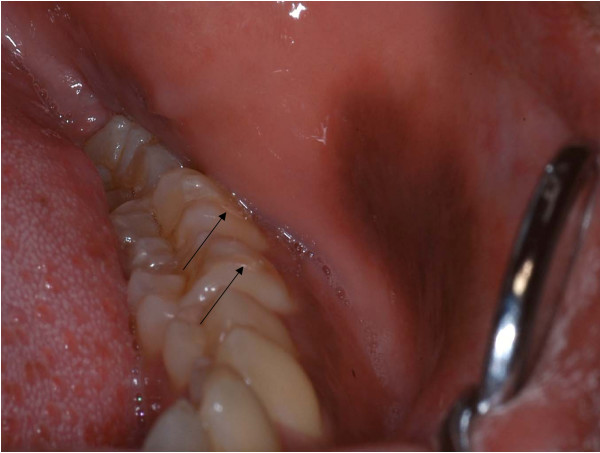
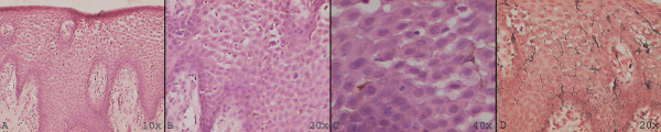
Case presentation: We present a case report of oral melanoacanthoma in a 24-year-old Asian Indian man. He presented with an intra-oral brown macular lesion on the left buccal mucosa with a duration of one and a half months. Microscopic examination revealed acanthosis of stratified squamous surface epithelium and dendritic melanocytes diffusely distributed in the epithelium; the Masson-Fontana silver impregnation technique was used to demonstrate the dendritic melanocytes. Based on the history, clinical features and histological presentation, the lesion was diagnosed as melanoacanthoma.
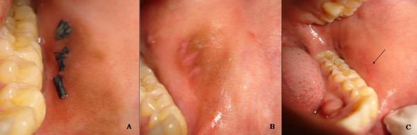
Conclusion: This is the first reported instance of oral melanoacanthoma in the Indian sub-continent. This report details the course of the lesion from diagnosis to its resolution. Melanoacanthoma must be differentiated from other intra-oral pigmented lesions and biopsy may be required to rule out melanoma.
Figures
Similar articles
-
An unusual clinical presentation of gingival melanoacanthoma.J Indian Soc Periodontol. 2013 Sep;17(5):657-60. doi: 10.4103/0972-124X.119288. J Indian Soc Periodontol. 2013. PMID: 24174763 Free PMC article.
-
A rare case of oral melanoacanthoma.J Oral Maxillofac Pathol. 2018 Sep-Dec;22(3):410-412. doi: 10.4103/jomfp.JOMFP_96_18. J Oral Maxillofac Pathol. 2018. PMID: 30651689 Free PMC article.
-
Oral melanoacanthoma of a rare intraoral site: case report and review of literature.Int J Clin Pediatr Dent. 2013 Jan;6(1):40-3. doi: 10.5005/jp-journals-10005-1185. Epub 2013 Apr 26. Int J Clin Pediatr Dent. 2013. PMID: 25206187 Free PMC article.
-
Oral Melanoacanthoma: Case Series of 33 Cases and Review of the Literature.Head Neck Pathol. 2023 Jun;17(2):364-370. doi: 10.1007/s12105-022-01506-w. Epub 2022 Dec 7. Head Neck Pathol. 2023. PMID: 36478543 Free PMC article. Review.
-
Solitary and multifocal oral melanoacanthoma.Int J Dermatol. 2007 Dec;46(12):1232-6. doi: 10.1111/j.1365-4632.2007.03393.x. Int J Dermatol. 2007. PMID: 18173514 Review.
Cited by
-
Immunohistochemical features of multifocal melanoacanthoma in the hard palate: a case report.BMC Res Notes. 2013 Jan 28;6:30. doi: 10.1186/1756-0500-6-30. BMC Res Notes. 2013. PMID: 23356913 Free PMC article.
-
Oral melanoacanthoma of the palate: An unusual presentation of an uncommon entity.JAAD Case Rep. 2018 Jan 16;4(2):138-139. doi: 10.1016/j.jdcr.2017.11.023. eCollection 2018 Mar. JAAD Case Rep. 2018. PMID: 29387765 Free PMC article. No abstract available.
-
Oral melanoacanthoma: A rare case of diffuse oral pigmentation.J Oral Maxillofac Pathol. 2012 Sep;16(3):441-3. doi: 10.4103/0973-029X.102514. J Oral Maxillofac Pathol. 2012. PMID: 23248484 Free PMC article.
-
Case report on trial: Do you, Doctor, swear to tell the truth, the whole truth and nothing but the truth?J Med Case Rep. 2011 May 13;5:179. doi: 10.1186/1752-1947-5-179. J Med Case Rep. 2011. PMID: 21569508 Free PMC article.
-
Mucosal Melanoma: Pathological Evolution, Pathway Dependency and Targeted Therapy.Front Oncol. 2021 Jul 19;11:702287. doi: 10.3389/fonc.2021.702287. eCollection 2021. Front Oncol. 2021. PMID: 34350118 Free PMC article. Review.
References
-
- Neville B, Damm DD, Allen CM, Bouquot J. Oral and Maxillofacial Pathology. Philadelphia, PA: WB Saunders Company; 2004.
LinkOut - more resources
Full Text Sources
