Review
. 2009 Jan 13:4:2.
doi: 10.1186/1750-1172-4-2.

Tetralogy of Fallot

Affiliations
Review

Tetralogy of Fallot

Frederique Bailliard et al. Orphanet J Rare Dis. .

Abstract

Tetralogy of Fallot is a congenital cardiac malformation that consists of an interventricular communication, also known as a ventricular septal defect, obstruction of the right ventricular outflow tract, override of the ventricular septum by the aortic root, and right ventricular hypertrophy. This combination of lesions occurs in 3 of every 10,000 live births, and accounts for 7-10% of all congenital cardiac malformations. Patients nowadays usually present as neonates, with cyanosis of varying intensity based on the degree of obstruction to flow of blood to the lungs. The aetiology is multifactorial, but reported associations include untreated maternal diabetes, phenylketonuria, and intake of retinoic acid. Associated chromosomal anomalies can include trisomies 21, 18, and 13, but recent experience points to the much more frequent association of microdeletions of chromosome 22. The risk of recurrence in families is 3%. Useful diagnostic tests are the chest radiograph, electrocardiogram, and echocardiogram. The echocardiogram establishes the definitive diagnosis, and usually provides sufficient information for planning of treatment, which is surgical. Approximately half of patients are now diagnosed antenatally. Differential diagnosis includes primary pulmonary causes of cyanosis, along with other cyanotic heart lesions, such as critical pulmonary stenosis and transposed arterial trunks. Neonates who present with ductal-dependent flow to the lungs will receive prostaglandins to maintain ductal patency until surgical intervention is performed. Initial intervention may be palliative, such as surgical creation of a systemic-to-pulmonary arterial shunt, but the trend in centres of excellence is increasingly towards neonatal complete repair. Centres that undertake neonatal palliation will perform the complete repair at the age of 4 to 6 months. Follow-up in patients born 30 years ago shows a rate of survival greater than 85%. Chronic issues that now face such adults include pulmonary regurgitation, recurrence of pulmonary stenosis, and ventricular arrhythmias. As the strategies for surgical and medical management have progressed, the morbidity and mortality of those born with tetralogy of Fallot in the current era is expected to be significantly improved.


Figures
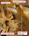
Figure 1
Figure 1
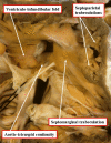
An autopsied specimen has been opened through the anterior wall of the right ventricle to show the cardinal features of tetralogy of Fallot.
Figure 2
Figure 2
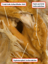
In this specimen, opened in the same fashion as the heart shown in Figure 1, there is fibrous continuity between the leaflets of the aortic and tricuspid valves in the postero-inferior margin of the ventricular septal defect, making the defect itself perimembranous.
Figure 3
Figure 3
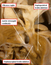
In this specimen, also photographed in a fashion comparable for the heart shown in Figure 1, there is muscular tissue interposed between the leaflets of the aortic and tricuspid valves in the postero-inferior margin of the ventricular septal defect.
Figure 4
Figure 4
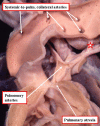
In this specimen, again photographed as for Figure 1, the defect extends to the level of the pulmonary valve due to failure of muscularisation of the outlet septum during development of the heart. This type of defect is doubly committed and juxtaarterial, but is also perimembranous.
Figure 5
Figure 5
This specimen has tetralogy of Fallot with pulmonary atresia. The pulmonary supply is through multiple systemic-to-pulmonary collateral arteries. The star shows the connection between one of the collateral arteries and the intrapericardial pulmonary arteries. All the other arteries join with the intrapericardial pulmonary arterial supply, or else supply segments of the lung directly. The task of the clinician is to display the supply of the various collateral arteries and their communications with the intrapericardial pulmonary arteries.
Figure 6
Figure 6
This still frame image of a parasternal short axis view of the echocardiogram of a patient with tetralogy of Fallot demonstrates the antero-cephalad deviation of the outlet septum into the right ventricular outflow tract.
Figure 7
Figure 7
A slightly modified view (a), angled to optimize imaging of the pulmonary arteries in the patient imaged to produce Figure 6, reveals significant hypoplasia of the pulmonary trunk and the pulmonary arteries, which result from the antero-cephalad deviation of the outlet septum. The pulmonary valvar leaflets are not visualized. In panel b, colour Doppler has been used, and demonstrates turbulence and acceleration of the flow of blood in the right ventricular outflow tract, originating at the level of the deviated outlet septum. The turbulence continues into the hypoplastic pulmonary trunk and pulmonary arteries.
Figure 8
Figure 8
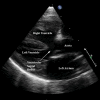
This still frame of a modified parasternal long axis view from the same patient as imaged for Figures 6 and 7 demonstrates the large ventricular septal defect, aortic override, and right ventricular hypertrophy charactistic of patients with tetralogy of Fallot.
Figure 9
Figure 9
Cardiac magnetic resonance imaging in an adult patient with tetralogy of Fallot repaired in childhood with an infundibular muscle resection and placement of a conduit from the right ventricle to the pulmonary arteries (a) has results in dilation and hypertrophy of the right ventricle. Additional images (b) show the stenotic and tortuous homograft conduit responsible for the dilated and hypertrophied right ventricle.

References

    1. Fallot ELA. Contribution a l'anatomie pathologique de la maladie bleu (cyanose cardiaque) Marseille Medical. pp. 77–93. - PubMed
    1. Becker AE, Connor M, Anderson RH. Tetralogy of Fallot: a morphometric and geometric study. American Journal of Cardiology. 1975;35:402–412. doi: 10.1016/0002-9149(75)90034-X. - DOI - PubMed
    1. Perry LW, Neill CA, Ferencz C, EUROCAT Working Party on. Congenital Heart Disease . Perspective in Pediatric Cardiology Epidemiology of congenital heart disease, the Baltimore-Washington Infant Study 1981–89. Armonk, NY: Futura; 1993. pp. 33–62.
    1. Oppenheimer-Dekker A, Bartelings MM, Wenink ACG. Anomalous architecture of the ventricles in hearts with overriding of aortic valve and a perimembranous ventricular septal defect ("Eisenmenger VSD") International Journal of Cardiology. 1989;9:341–355. doi: 10.1016/0167-5273(85)90032-4. - DOI - PubMed
    1. Anderson RH, Weinberg PM. The clinical anatomy of tetralogy of Fallot. Cardiology in the Young. 2005;15:38–47. doi: 10.1017/S1047951105001010. - DOI - PubMed