. 2009 Apr;83(7):3150-61.
doi: 10.1128/JVI.01456-08. Epub 2009 Jan 14.

Mengovirus-induced rearrangement of the nuclear pore complex: hijacking cellular phosphorylation machinery

Affiliations

Mengovirus-induced rearrangement of the nuclear pore complex: hijacking cellular phosphorylation machinery

Maryana V Bardina et al. J Virol. 2009 Apr.

Abstract

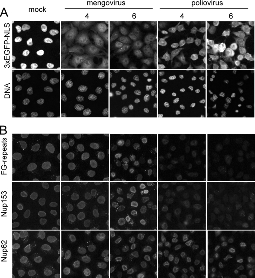
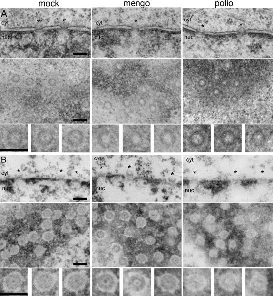
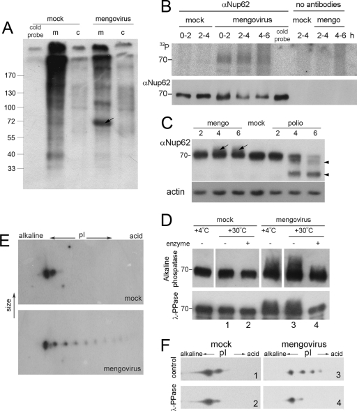
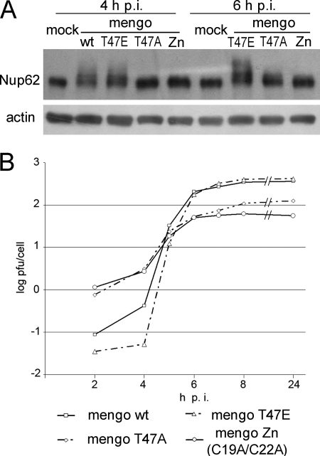
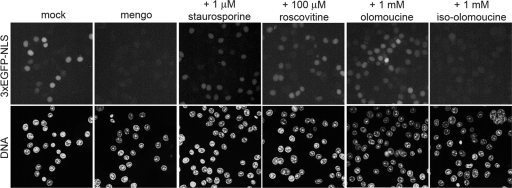
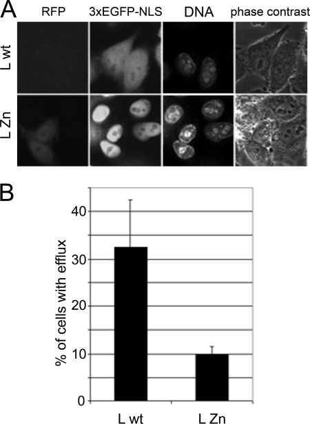
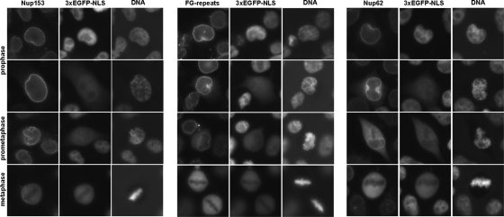
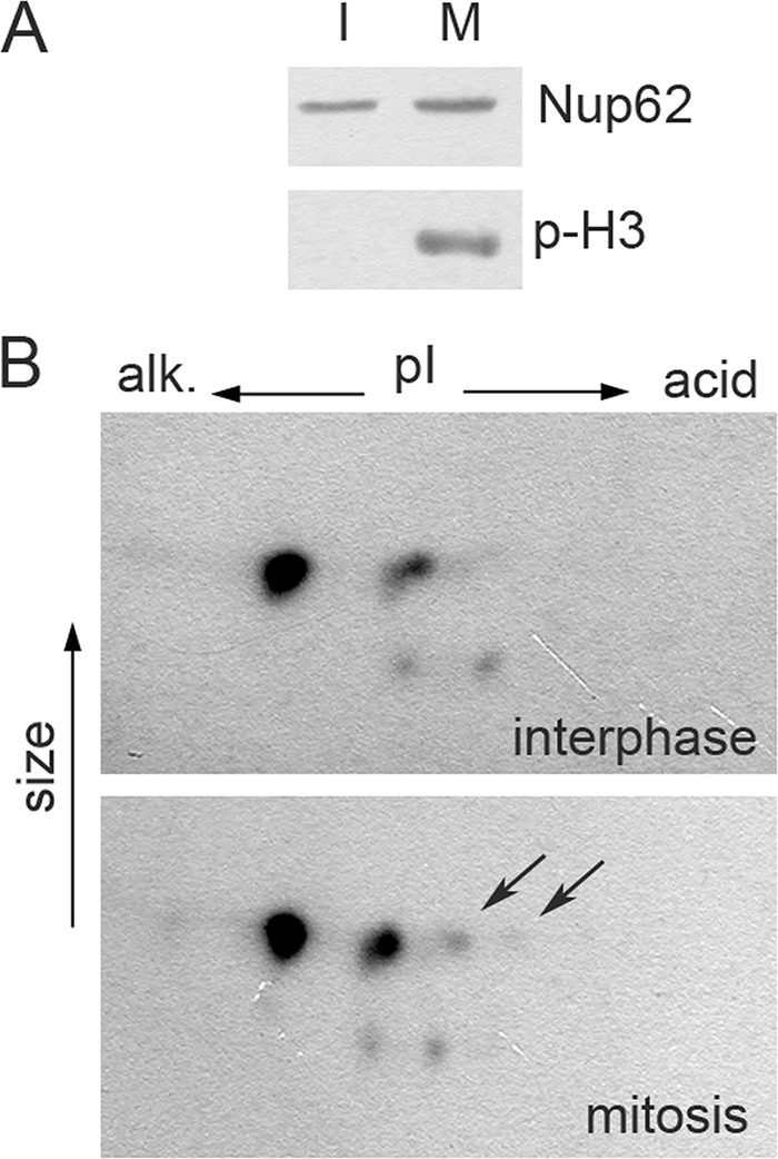
Representatives of several picornavirus genera have been shown previously to significantly enhance non-controllable bidirectional exchange of proteins between nuclei and cytoplasm. In enteroviruses and rhinoviruses, enhanced permeabilization of the nuclear pores appears to be primarily due to proteolytic degradation of some nucleoporins (protein components of the pore), whereas this effect in cardiovirus-infected cells is triggered by the leader (L) protein, devoid of any enzymatic activities. Here, we present evidence that expression of L alone was sufficient to cause permeabilization of the nuclear envelope in HeLa cells. In contrast to poliovirus, mengovirus infection of these cells did not elicit loss of nucleoporins Nup62 and Nup153 from the nuclear pore complex. Instead, nuclear envelope permeabilization was accompanied by hyperphosphorylation of Nup62 in cells infected with wild-type mengovirus, whereas both of these alterations were suppressed in L-deficient virus mutants. Since phosphorylation of Nup62 (although less prominent) did accompany permeabilization of the nuclear envelope prior to its mitotic disassembly in uninfected cells, we hypothesize that cardiovirus L alters the nucleocytoplasmic traffic by hijacking some components of the normal cell division machinery. The variability and biological significance of picornaviral interactions with the nucleocytoplasmic transport in the infected cells are discussed.


Figures

FIG. 1.
FIG. 1.
Localization of Nups in cells infected with mengovirus and poliovirus. HeLa-3E cells were infected with the viruses with an input MOI of ∼100 PFU/cell and fixed at indicated times. (A) Exit of the nuclear marker protein into the cytoplasm. (B) Detection of FG-repeat-containing proteins, Nup153, and Nup62 by immunostaining. Images in a given row were made with equal expositions.
FIG. 2.
FIG. 2.
Electron microscopy of nuclear pores in infected cells. HeLa-3E cells were infected with mengovirus and poliovirus and fixed at 4 h p.i. The upper rows in panels A and B correspond to cross-sectioned nuclear envelopes, the middle rows are tangentially cut nuclear envelopes in plane, and the lower rows represent the twofold-enlarged examples of NPC from the appropriate middle rows. Scales bars correspond to 200 nm. The nuclear sides in cross-sections are marked as “nuc,” and the cytoplasmic ones are marked as “cyt.” Regions of NPC are marked with asterisks. (A) In situ fixation. The bar-like structures visible in NPC cross-sections of healthy cells are absent from a number of pores after poliovirus and mengovirus infections. In tangential section, control NPC display an electron-dense granule in the central channel, which is totally or partially lost in infected cells. (B) Fixation after dissolution of membranes with Triton X-100. NPC attached to the lamina are visible in cross-sections of the control envelope, and these complexes appeared to be only slightly disorganized in the mengovirus-infected sample. On the other hand, they are significantly destroyed by poliovirus infection. In tangential sections, the pores in control and mengovirus-infected cells looked very similar, whereas the NPC from poliovirus-infected cells displayed a much higher electron transparency and are less structured.
FIG. 3.
FIG. 3.
Nup62 is hyperphosphorylated in mengovirus-infected cells. (A) [32P]phosphate incorporation in WGA-binding proteins upon cardiovirus infection. HeLa-3E cells were labeled with [32P]orthophosphate during the first 2 h of infection, lysed, and subfractionated into cytosolic (c) and membrane (m) fractions as described in Materials and Methods. Nups were precipitated with WGA-beads and analyzed by PAGE and autoradiography. The first lane represents the control probe incubated for 2 h in ice. The arrow points out to a ∼70-kDa band appearing in mengovirus-infected cells. (B) Immunoprecipitation of 32P-labeled Nup62 from mengovirus-infected cells. Infected and mock-infected HeLa-3E cells were labeled with inorganic [32P]phosphate during the time periods indicated. The cold probe was exposed to the label for 6 h in ice. The cells were lysed and Nup62 was immunoprecipitated and subjected to electrophoresis as described in Materials and Methods (upper row). The amounts of Nup62 in the samples were controlled by Western blotting (lower row). (C) Modifications of Nup62 by different picornaviruses. HeLa-3E cells were infected with mengovirus or poliovirus for indicated times, and their extracts were investigated by PAGE, followed by Western blotting with anti-Nup62 antibodies. The lower mobility component of Nup62 from mengovirus-infected cells are marked with arrows; higher-mobility bands from poliovirus-infected cells corresponding to degradation products are marked with triangles. (D) The low-mobility forms of Nup62 disappeared from mengovirus-infected cell after alkaline phosphatase (upper row) and λ-PPase (lower row) treatments. Lysates from mock- and mengovirus-infected cells made at 4 h p.i. in the case of the former enzyme and immunoprecipitated preparations of Nup62 from such lysates in the case of the latter were treated with the phosphatases for 30 min at 30°C and analyzed by Western blotting. (E) Detection of hyperphosphorylated forms of Nup62 in mengovirus-infected cells. Cell lysates were prepared as in panel C and were subjected to 2D electrophoresis followed by Western blotting. (F) The additional acidic spots of Nup62 disappeared after treatment with λ-PPase. Samples 1 to 4 from the lower row of panel D were subjected to 2D electrophoresis, followed by Western blotting.
FIG. 4.
FIG. 4.
Effect of L protein mutations on Nup62 phosphorylation. (A) HeLa-3E cells were infected with wild-type (wt) and T47E mengoviruses at an MOI of ∼20 PFU/cell and with the C19A/C22A (Zn) and T47A mutants at an MOI of ∼100 PFU/cell to compensate for their relative replicative deficiencies. After incubation, the cells were observed under epifluorescence microscope (results not shown) and harvested for Western blotting and titration. (B) Growth curves of mengovirus mutants at the MOIs indicated above.
FIG. 5.
FIG. 5.
Mengovirus-induced permeabilization of the nuclear envelope is sensitive to protein kinase inhibitors. Permeabilized uninfected HeLa-3E cells were incubated with the lysates from mengovirus-infected (4 h p.i.) and mock-infected HeLa cells for 1 h at 37°C in the absence or presence of the indicated concentrations of a broad-spectrum protein kinase inhibitor staurosporin and two inhibitors of cycle-dependent kinases, roscovitine and olomoucine. An inactive analog of the latter drug, iso-olomoucine, was used as a control.
FIG. 6.
FIG. 6.
Individually expressed leader protein affects nucleocytoplasmic traffic. HeLa-3E cells were transfected with the plasmids encoding leader proteins fused to RFP. (A) Efflux of the nuclear protein marker (3×EGFP-NLS) into the cytoplasm is visible in a proportion of cells transfected with the plasmid encoding wild-type (wt) L protein but not with the one encoding inactive C19A/C22A (Zn) mutant. Cells were stained with Hoechst 33342 DNA stain and captured 24 h posttransfection. (B) Percentage of the cells transfected with wild-type (wt) and mutant L-expressing plasmids, demonstrating 3×EGFP-NLS fluorescence in the cytoplasm. The transfected cells were incubated in the presence of 100 μM zVAD(OMe).fmk to prevent apoptosis, which could complicate interpretation of the results.
FIG. 7.
FIG. 7.
Localization of Nups and efflux of the nuclear protein marker into cytoplasm in prophase and metaphase cells. Uninfected HeLa-3E cells at different stages of mitosis, as judged by DNA staining, were analyzed with respect to localization of Nups and distribution of the fluorescent marker. Prophase cells displaying 3×EGFP-NLS in cytoplasm possessed Nups mainly associated with the nuclear envelope.
FIG. 8.
FIG. 8.
Nup62 is phosphorylated in mitotic HeLa cells. (A) Control for synchronization. Samples of interphase (I) and mitotic (M) cells (see Materials and Methods) were analyzed by Western blotting for Nup62 and Ser-10 phosphorylated H3 histone (p-H3). (B) The same preparations were analyzed by 2D electrophoresis, followed by Western blotting for Nup62. Acidic spots are visible in the preparations of mitotic cells, but those of interphase cells are marked by arrows.

References

    1. Adam, S. A., S. R. Marr, and L. Gerace. 1990. Nuclear protein import in permeabilized mammalian cells requires soluble cytoplasmic factors. J. Cell Biol. 111807-816. - PMC - PubMed
    1. Agol, V. I. 2002. Picornavirus genome: an overview, p. 127-148. In B. L. Semler and E. Wimmer (ed.), Molecular biology of picornaviruses. ASM Press, Washington, DC.
    1. Almstead, L. L., and P. Sarnow. 2007. Inhibition of U snRNP assembly by a virus-encoded proteinase. Genes Dev. 211086-1097. - PMC - PubMed
    1. Beck, M., F. Förster, M. Ecke, J. M. Plitzko, F. Melchior, G. Gerisch, W. Baumeister, and O. Medalia. 2004. Nuclear pore complex structure and dynamics revealed by cryoelectron tomography. Science 3061387-1390. - PubMed
    1. Belov, G. A., A. G. Evstafieva, O. V. Mikitas, A. B. Vartapetian, and V. I. Agol. 2000. Early alteration of nucleocytoplasmic traffic induced by some RNA viruses. Virology 275244-248. - PubMed

Publication types

Substances