. 2009 Jan 14;29(2):444-53.
doi: 10.1523/JNEUROSCI.4029-08.2009.

Single nigrostriatal dopaminergic neurons form widely spread and highly dense axonal arborizations in the neostriatum

Affiliations

Single nigrostriatal dopaminergic neurons form widely spread and highly dense axonal arborizations in the neostriatum

Wakoto Matsuda et al. J Neurosci. .

Abstract

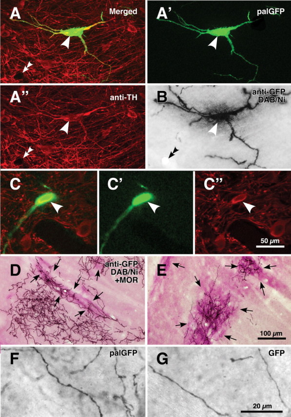
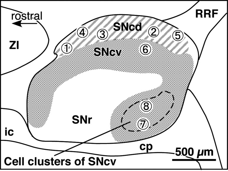
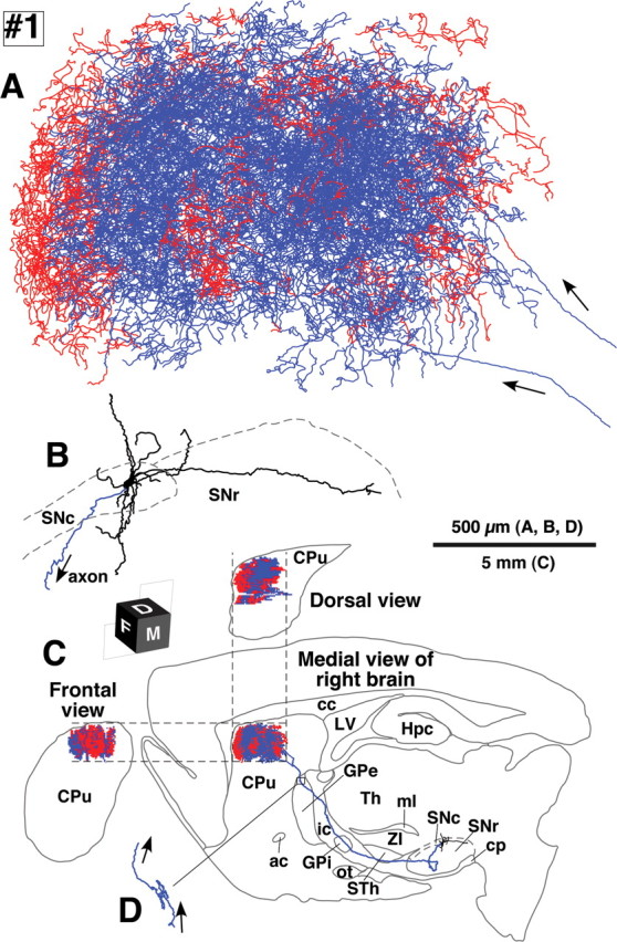
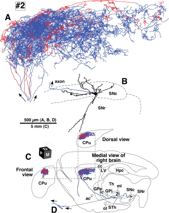
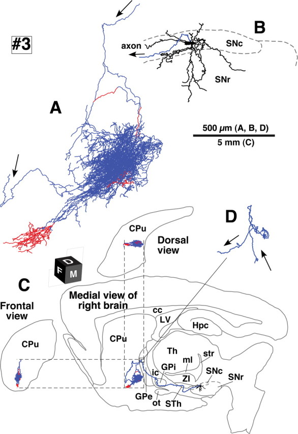
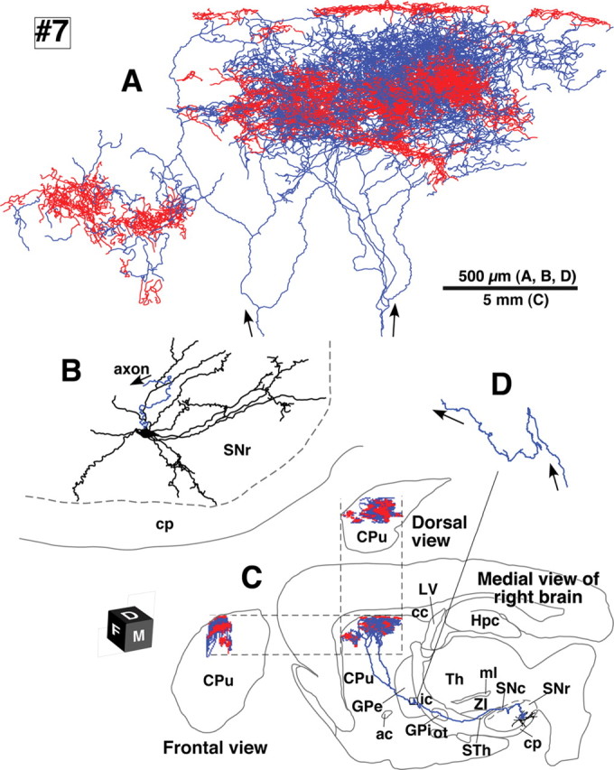
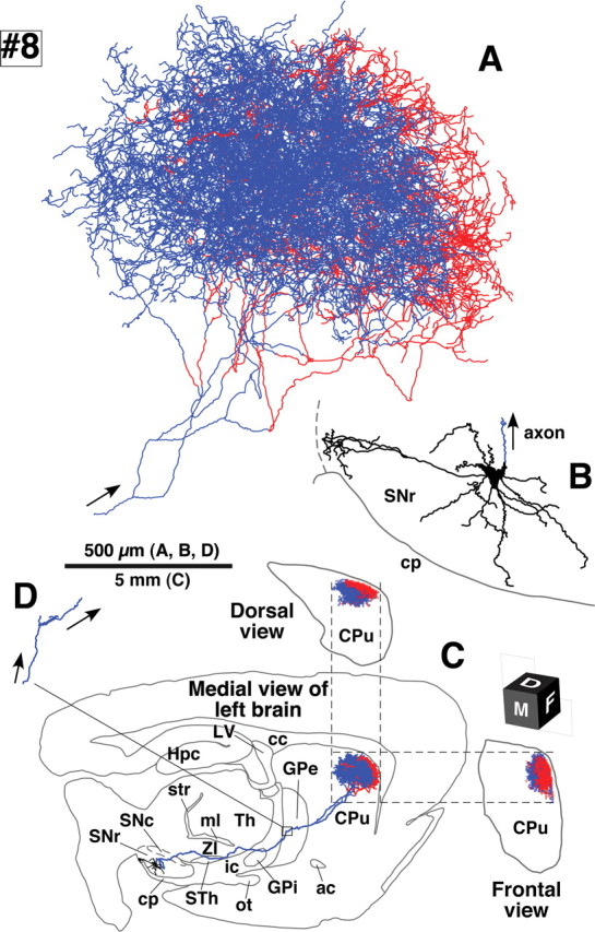
The axonal arbors of single nigrostriatal dopaminergic neurons were visualized with a viral vector expressing membrane-targeted green fluorescent protein in rat brain. All eight reconstructed tyrosine hydroxylase-positive dopaminergic neurons possessed widely spread and highly dense axonal arborizations in the neostriatum. All of them emitted very little axon collateral arborization outside of the striatum except for tiny arborization in the external pallidum. The striatal axonal bush of each reconstructed dopaminergic neuron covered 0.45-5.7% (mean +/- SD = 2.7 +/- 1.5%) of the total volume of the neostriatum. Furthermore, all the dopaminergic neurons innervated both striosome and matrix compartments of the neostriatum, although each neuron's arborization tended to favor one of these compartments. Our findings demonstrate that individual dopaminergic neurons of the substantia nigra can broadcast a dopamine signal and exert strong influence over a large number of striatal neurons. This divergent signaling should be a key to the function of the nigrostriatal system in dopamine-based learning and suggests that neurodegeneration of individual nigral neurons can affect multiple neurons in the striatum. Thus, these results would also contribute to understanding the clinicopathology of Parkinson's disease and related syndromes.


Figures

Figure 1.
Figure 1.
Single-neuron labeling with palGFP Sindbis virus vector. A nigral neuron showing green fluorescence of palGFP (A, A') was immunolabeled for TH (A, A“), and further stained black by the immunoperoxidase method with anti-GFP antibody and diaminobenzidine/nickel reaction (DAB/Ni) (B). Double arrowheads in A and B point to the same small vessel. Another example for palGFP-labeled TH-positive neurons is indicated by arrowheads in C–C”. The black-stained axon fibers of palGFP-labeled neurons densely innervated the striatum (D, E), being distributed both in the striosome/patch and matrix compartments (D) or with preference for the striosome compartment (E). MOR immunoreactivity was used to label the striosome compartments (violet–red areas indicated by arrows in D, E). palGFP-labeled axon fibers (F) were compared with GFP-labeled fibers (G) at a higher magnification. Scale bar in C“ applies to A–C″, that in E applies to D, E, and that in G applies to F, G.
Figure 2.
Figure 2.
The distribution of the eight neurons labeled with palGFP Sindbis virus vector and immunopositive for tyrosine hydroxylase in the substantia nigra. The locations were projected onto a parasagittal plane 2.25 mm lateral to the midline. Each encircled numeral indicates the position of the neuron with the same number in Table 1. cp, Cerebral peduncle; ic, internal capsule; ZI, zona incerta.
Figure 3.
Figure 3.
Camera lucida reconstruction of SNcd neuron #1. A, B, The axon fibers in the striatum (A) and dendrites (B) in the SNc were projected onto a parasagittal plane and superimposed from the medial side. C, The dorsal and frontal views of the intrastriatal axonal arborization were reconstructed and compared with the medial view. Red and blue lines in the striatum indicate the axon fibers located in the striosome and matrix compartments, respectively. Red fibers at the most rostral portion in A were mostly located in the MOR-positive subcallosal streak. D, The axon gave rise to only minor collaterals in the external segment of the GPe. ac, Anterior commissure; cc, corpus callosum; cp, cerebral peduncle; CPu, caudate–putamen (neostriatum); Hpc, hippocampus; ic, internal capsule; LV, lateral ventricle; ml, medial lemniscus; ot, optic tract; STh, subthalamic nucleus; str, superior thalamic radiation; Th, thalamus; ZI, zona incerta.
Figure 4.
Figure 4.
Camera lucida reconstruction of SNcd neuron #2. This neuron did not emit axon collateral in the GPe, but took a loop-making meandering course through the subthalamic nucleus (STh; D). Main axons meandering in the STh were found only in neurons #2 and #4. See the legend of Figure 3 for further detail.
Figure 5.
Figure 5.
Camera lucida reconstruction of SNcd neuron #3. Of the eight neurons examined in the present study, this neuron had the smallest axonal arborization in the striatum. See the legend of Figure 3 for further detail.
Figure 6.
Figure 6.
Camera lucida reconstruction of SNcv neuron #7. The axons of this SNcv neuron showed the highest preference for the striosome compartment in the present results and made two bushes in the neostriatum. Red fibers at the top of A were located in MOR-positive subcallosal streaks. See the legend of Figure 3 for further detail.
Figure 7.
Figure 7.
Camera lucida reconstruction of SNcv neuron #8. Although the cell body of this neuron was located at the cell cluster region of the SNcv, the axonal arborization was very similar to that of SNcd neuron #1 (Fig. 3). See the legend of Figure 3 for detail.

References

    1. Alexander GE, Crutcher MD, DeLong MR. Basal ganglia-thalamocortical circuits: parallel substrates for motor, oculomotor, “prefrontal” and “limbic” functions. Prog Brain Res. 1990;85:119–146. - PubMed
    1. Andén NE, Hfuxe K, Hamberger B, Hökfelt T. A quantitative study on the nigro-neostriatal dopamine neuron system in the rat. Acta Physiol Scand. 1966;67:306–312. - PubMed
    1. Arluison M, Dietl M, Thibault J. Ultrastructural morphology of dopaminergic nerve terminals and synapses in the striatum of the rat using tyrosine hydroxylase immunocytochemistry: a topographical study. Brain Res Bull. 1984;13:269–285. - PubMed
    1. Björklund A, Dunnett SB. Dopamine neuron systems in the brain: an update. Trends Neurosci. 2007;30:194–202. - PubMed
    1. Bowen WD, Gentleman S, Herkenham M, Pert CB. Interconverting mu and delta forms of the opiate receptor in rat striatal patches. Proc Natl Acad Sci U S A. 1981;78:4818–4822. - PMC - PubMed

Publication types