. 2009 Mar;234(3):354-60.
doi: 10.3181/0806-BC-194. Epub 2009 Jan 14.

Enhanced CD36 scavenger receptor expression in THP-1 human monocytes in the presence of lupus plasma: linking autoimmunity and atherosclerosis

Affiliations

Enhanced CD36 scavenger receptor expression in THP-1 human monocytes in the presence of lupus plasma: linking autoimmunity and atherosclerosis

Allison B Reiss et al. Exp Biol Med (Maywood). 2009 Mar.

Abstract

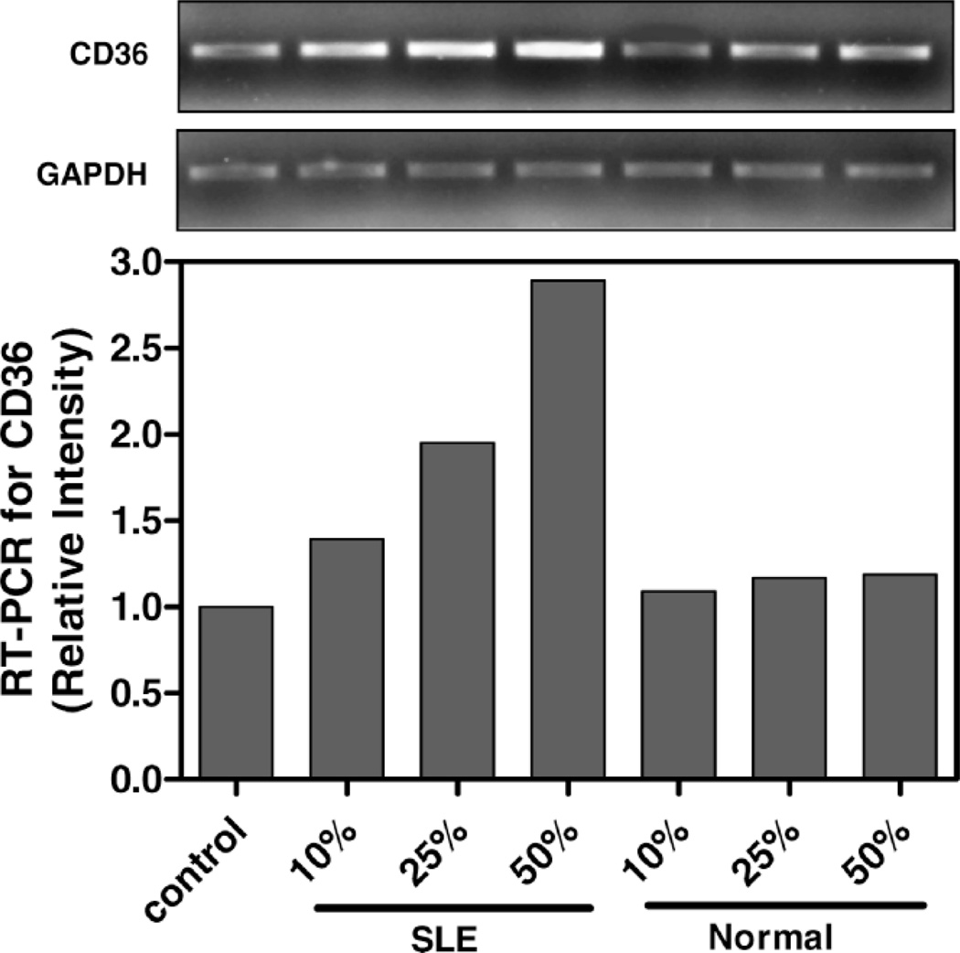
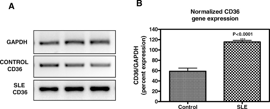
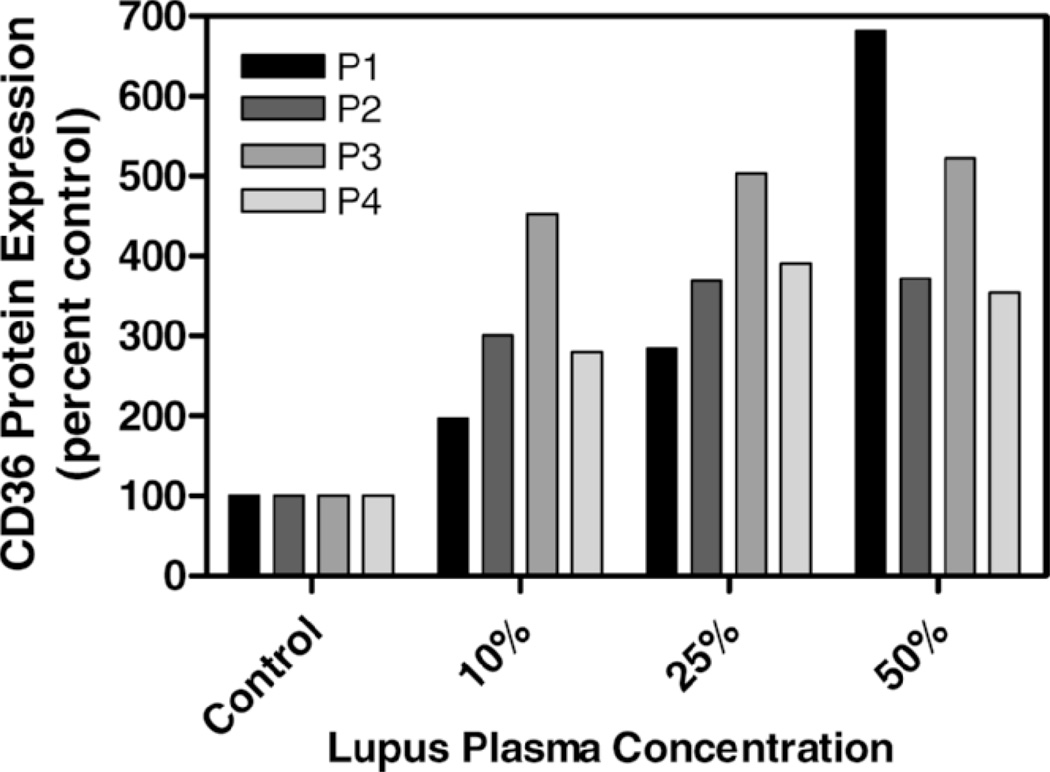
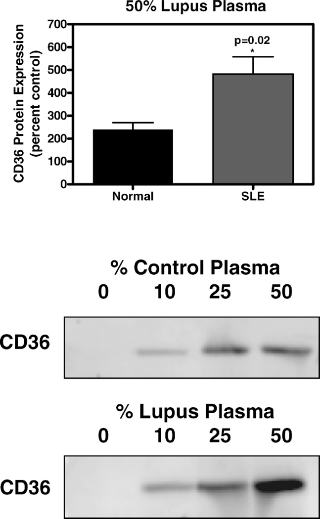
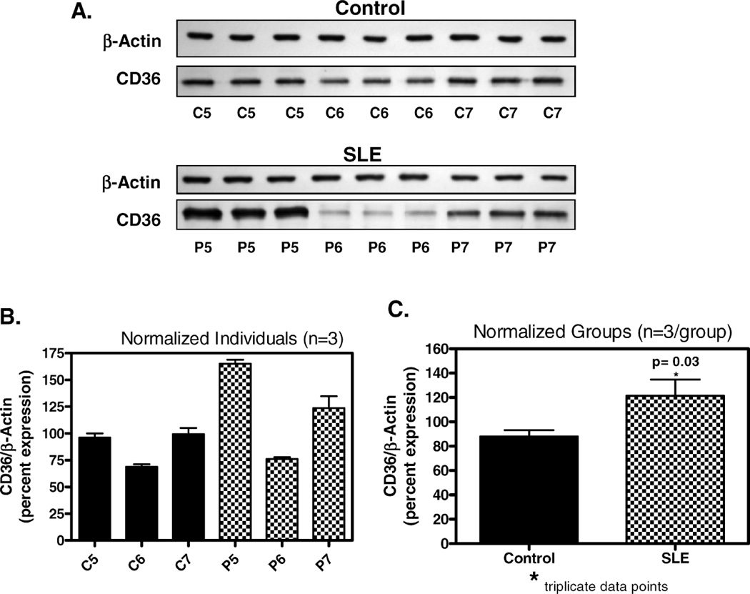
Premature atherosclerotic cardiovascular disease (ASCVD) is a common and devastating complication of systemic lupus erythematosus (SLE). It is likely that immunologic derangements contribute to premature ASCVD in these patients, possibly by disrupting homeostatic mechanisms that orchestrate cholesterol balance in monocytes/macrophages in the artery wall. CD36, a macrophage scavenger receptor responsible for recognition and internalization of oxidized lipids, is a major participant in atherosclerotic foam cell formation. We hypothesized that lupus plasma would affect CD36 expression in a pro-atherogenic manner in THP-1 human monocytes and differentiated macrophages. SLE patient plasma markedly stimulated expression of CD36 message in a dose-dependent fashion in THP-1 human monocytes. A 50% volume/volume concentration of plasma derived from SLE patients increased CD36 mRNA by 71 +/- 8% (n = 3, P < 0.001) above 50% normal human plasma. 50% SLE patient plasma increased CD36 mRNA expression to 290 +/- 12% of no-plasma control (n = 3, P < 0.001), compared with only 118 +/- 3.7% of control in the presence of 50% normal human plasma (n = 3, not significant). 50% lupus plasma also upregulated CD36 protein expression by 482.3 +/- 76.2% (n = 4, P < 0.05), whereas the presence of 50% normal human plasma increased the CD36 protein level by only 239.8 +/- 61.9% (n = 4, P < 0.05). To our knowledge, this is the first demonstration that CD36 expression is enhanced by plasma from patients with an autoimmune disorder. Premature atherosclerosis is common in SLE patients. Upregulation of CD36 may contribute to this pathological process by increasing vulnerability to cholesterol overload. Demonstration of disrupted cholesterol homeostasis in this select group of patients provides further evidence of the involvement of the immune system in atherogenesis and may inform us of the role of CD36 in the general atherogenic process. CD36 may provide a novel therapeutic target in the treatment of ASCVD in SLE patients.


Figures

Figure 1
Figure 1
CD36 message level in THP-1 monocytes increase with SLE plasma exposure. THP-1 monocytes were incubated in increasing concentrations of SLE patient or normal control plasma for 3 hours as indicated. Total RNA isolated from cells exposed to each condition was reverse transcribed and amplified by PCR with GAPDH message as an internal standard. Representative experiment from a total of three SLE patients studied.
Figure 2
Figure 2
Effect of SLE plasma versus normal human plasma on CD36 mRNA in THP-1 monocytes. THP-1 monocytes were incubated in 50% volume/volume SLE patient or normal control plasma for 3 hours as indicated. Following incubation, total RNA isolated from cells exposed to each condition was reverse transcribed and amplified by PCR with GAPDH message as an internal standard. A) Representative experiment from a total of three SLE patients and three controls studied. Photograph of ethidium bromide-stained PCR-amplified bands corresponding to message for CD36 and GAPDH as indicated. B) Gene expression levels were graphed as relative mRNA expression. The data represent the mean and SEM of three independent experiments.
Figure 3
Figure 3
Effect of plasma from four individual SLE patients on CD36 protein in THP-1 monocytes. Cultured THP-1 cells were untreated (control, 0% plasma) or exposed for 6 hours to increasing concentrations of plasma from 4 distinct SLE patients. Total cellular protein was isolated and run on an SDS-polyacrylamide gel and immunoblotted with mouse monoclonal IgM anti-peptide antibody raised against human CD36 protein.
Figure 4
Figure 4
CD36 protein expression in THP-1 monocytes exposed to SLE plasma and healthy control plasma. Cultured THP-1 monocytes were exposed for 6 hours to SLE or control plasma as indicated. Total cellular protein was isolated and run on an SDS-polyacrylamide gel and immunoblotted with mouse monoclonal IgM anti-peptide antibody raised against human CD36 protein. Upper portion of figure is a graphic representation of the difference in CD36 protein level in cells incubated for 12 hours in SLE patient plasma versus control plasma (n = 4 for each). Lower portion of figure shows marked increase in CD36 band intensity with increasing percentage of plasma from a single representative SLE patient versus a single control normal human plasma patient.
Figure 5
Figure 5
Effect of SLE plasma on CD36 protein in THP-1 monocytes. Cultured THP-1 monocytes were exposed for 12 hours to SLE or control plasma as indicated. Total cellular protein was isolated and run on an SDS-polyacrylamide gel and immunoblotted with mouse monoclonal IgM anti-peptide antibody raised against human CD36 protein. A) CD36 expression in 3 control subjects with corresponding beta-actins (C5, C6, C7) compared to 3 SLE patients (P5, P6, P7) shows elevated CD36 expression in 2 of the 3 SLE patients. B) Graphic representation of immunoblot results from section A with normalization to beta-actin. C) Combined data from Figure B representing aggregate of controls versus patients for this experiment, normalized to beta-actin with statistically significant difference in CD36 expression.

References

    1. Asanuma Y, Oeser A, Shintani AK, Turner E, Olsen N, Fazio S, Linton MF, Raggi P, Stein CM. Premature coronary-artery atherosclerosis in systemic lupus erythematosus. N Engl J Med. 2003;349:2407–2415. - PubMed
    1. Hahn B, McMahon M. Atherosclerosis and systemic lupus erythematosus: the role of altered lipids and of autoantibodies. Lupus. 2008;17:368–370. - PubMed
    1. Frostegard J. Systemic lupus erythematosus and cardiovascular disease. Lupus. 2008;17:364–367. - PubMed
    1. Esdaile JM, Panaritis C, Abrahamowicz M. Traditional Framingham risk factors fail to fully account for accelerated atherosclerosis in systemic lupus erythematosus. Arthritis Rheum. 2001;44:2331–2337. - PubMed
    1. McMahon M, Hahn B. Atherosclerosis and systemic lupus erythematosus: mechanistic basis of the association. Curr Opin Immunol. 2007;19:633–639. - PMC - PubMed

Publication types