

Coarse-grained modeling of large RNA molecules with knowledge-based potentials and structural filters
- PMID: 19144906
- PMCID: PMC2648710
- DOI: 10.1261/rna.1270809
Coarse-grained modeling of large RNA molecules with knowledge-based potentials and structural filters
Abstract
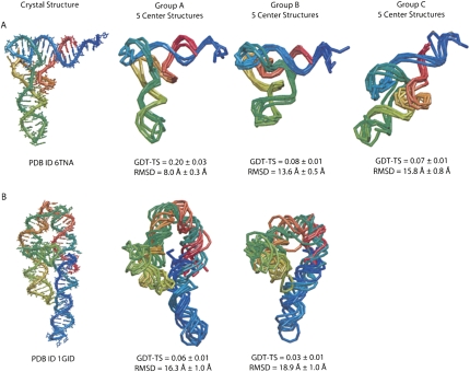
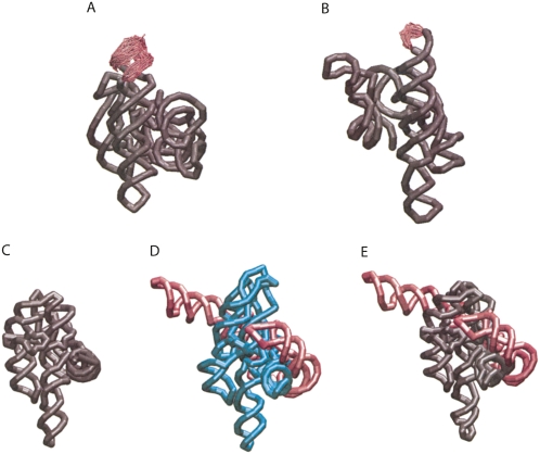
Understanding the function of complex RNA molecules depends critically on understanding their structure. However, creating three-dimensional (3D) structural models of RNA remains a significant challenge. We present a protocol (the nucleic acid simulation tool [NAST]) for RNA modeling that uses an RNA-specific knowledge-based potential in a coarse-grained molecular dynamics engine to generate plausible 3D structures. We demonstrate NAST's capabilities by using only secondary structure and tertiary contact predictions to generate, cluster, and rank structures. Representative structures in the best ranking clusters averaged 8.0 +/- 0.3 A and 16.3 +/- 1.0 A RMSD for the yeast phenylalanine tRNA and the P4-P6 domain of the Tetrahymena thermophila group I intron, respectively. The coarse-grained resolution allows us to model large molecules such as the 158-residue P4-P6 or the 388-residue T. thermophila group I intron. One advantage of NAST is the ability to rank clusters of structurally similar decoys based on their compatibility with experimental data. We successfully used ideal small-angle X-ray scattering data and both ideal and experimental solvent accessibility data to select the best cluster of structures for both tRNA and P4-P6. Finally, we used NAST to build in missing loops in the crystal structures of the Azoarcus and Twort ribozymes, and to incorporate crystallographic data into the Michel-Westhof model of the T. thermophila group I intron, creating an integrated model of the entire molecule. Our software package is freely available at https://simtk.org/home/nast.
Figures
Similar articles
-
Predicting RNA structure by multiple template homology modeling.Pac Symp Biocomput. 2010:216-27. doi: 10.1142/9789814295291_0024. Pac Symp Biocomput. 2010. PMID: 19908374 Free PMC article.
-
RNA structure determination using SAXS data.J Phys Chem B. 2010 Aug 12;114(31):10039-48. doi: 10.1021/jp1057308. J Phys Chem B. 2010. PMID: 20684627 Free PMC article.
-
Automated RNA tertiary structure prediction from secondary structure and low-resolution restraints.J Comput Chem. 2011 Jul 30;32(10):2232-44. doi: 10.1002/jcc.21806. Epub 2011 Apr 21. J Comput Chem. 2011. PMID: 21509787 Free PMC article.
-
RNA tectonics: towards RNA design.Fold Des. 1996;1(4):R78-88. doi: 10.1016/S1359-0278(96)00037-5. Fold Des. 1996. PMID: 9079386 Review.
-
Mapping nucleic acid structure by hydroxyl radical cleavage.Curr Opin Chem Biol. 2005 Apr;9(2):127-34. doi: 10.1016/j.cbpa.2005.02.009. Curr Opin Chem Biol. 2005. PMID: 15811796 Review.
Cited by
-
Elastic network models for RNA: a comparative assessment with molecular dynamics and SHAPE experiments.Nucleic Acids Res. 2015 Sep 3;43(15):7260-9. doi: 10.1093/nar/gkv708. Epub 2015 Jul 17. Nucleic Acids Res. 2015. PMID: 26187990 Free PMC article.
-
Quantifying the relationship between sequence and three-dimensional structure conservation in RNA.BMC Bioinformatics. 2010 Jun 15;11:322. doi: 10.1186/1471-2105-11-322. BMC Bioinformatics. 2010. PMID: 20550657 Free PMC article.
-
Limits in accuracy and a strategy of RNA structure prediction using experimental information.Nucleic Acids Res. 2019 Jun 20;47(11):5563-5572. doi: 10.1093/nar/gkz427. Nucleic Acids Res. 2019. PMID: 31106330 Free PMC article.
-
State-of-the-RNArt: benchmarking current methods for RNA 3D structure prediction.NAR Genom Bioinform. 2024 May 14;6(2):lqae048. doi: 10.1093/nargab/lqae048. eCollection 2024 Jun. NAR Genom Bioinform. 2024. PMID: 38745991 Free PMC article.
-
RNAlyzer--novel approach for quality analysis of RNA structural models.Nucleic Acids Res. 2013 Jul;41(12):5978-90. doi: 10.1093/nar/gkt318. Epub 2013 Apr 25. Nucleic Acids Res. 2013. PMID: 23620294 Free PMC article.
References
-
- Baker D., Sali A. Protein structure prediction and structural genomics. Science. 2001;294:93–96. - PubMed
-
- Ban N., Nissen P., Hansen J., Moore P.B., Steitz T.A. The complete atomic structure of the large ribosomal subunit at 2.4 Å resolution. Science. 2000;289:905–920. - PubMed
-
- Cate J.H., Gooding A.R., Podell E., Zhou K., Golden B.L., Kundrot C.E., Cech T.R., Doudna J.A. Crystal structure of a group I ribozyme domain: Principles of RNA packing. Science. 1996;273:1678–1685. - PubMed
Publication types
MeSH terms
Substances
Grants and funding
LinkOut - more resources
Full Text Sources
Molecular Biology Databases
Research Materials