

Genetic and structural analysis of base substitutions in the central pseudoknot of Thermus thermophilus 16S ribosomal RNA
- PMID: 19144908
- PMCID: PMC2648708
- DOI: 10.1261/rna.1374809
Genetic and structural analysis of base substitutions in the central pseudoknot of Thermus thermophilus 16S ribosomal RNA
Abstract
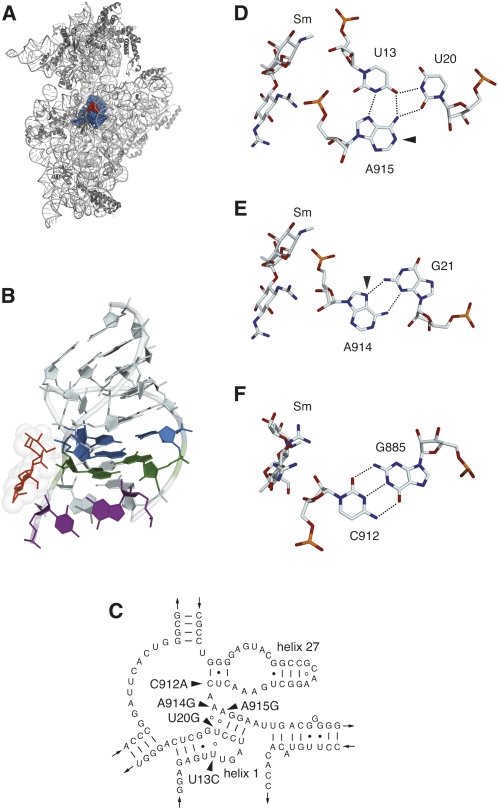
Characterization of base substitutions in rRNAs has provided important insights into the mechanism of protein synthesis. Knowledge of the structural effects of such alterations is limited, and could be greatly expanded with the development of a genetic system based on an organism amenable to both genetics and structural biology. Here, we describe the genetic analysis of base substitutions in 16S ribosomal RNA of the extreme thermophile Thermus thermophilus, and an analysis of the conformational effects of these substitutions by structure probing with base-specific modifying agents. Gene replacement methods were used to construct a derivative of strain HB8 carrying a single 16S rRNA gene, allowing the isolation of spontaneous streptomycin-resistant mutants and subsequent genetic mapping of mutations by recombination. The residues altered to give streptomycin resistance reside within the central pseudoknot structure of 16S rRNA comprised of helices 1 and 27, and participate in the U13-U20-A915 base triple, the G21-A914 type II sheared G-A base pair, or the G885-C912 Watson-Crick base pair closing helix 27. Substitutions at any of the three residues engaged in the base triple were found to confer resistance. Results from structure probing of the pseudoknot are consistent with perturbation of RNA conformation by these substitutions, potentially explaining their streptomycin-resistance phenotypes.
Figures
Similar articles
-
A mutation in the decoding center of Thermus thermophilus 16S rRNA suggests a novel mechanism of streptomycin resistance.J Bacteriol. 2005 Mar;187(6):2200-2. doi: 10.1128/JB.187.6.2200-2202.2005. J Bacteriol. 2005. PMID: 15743969 Free PMC article.
-
Structural analysis of base substitutions in Thermus thermophilus 16S rRNA conferring streptomycin resistance.Antimicrob Agents Chemother. 2014 Aug;58(8):4308-17. doi: 10.1128/AAC.02857-14. Epub 2014 May 12. Antimicrob Agents Chemother. 2014. PMID: 24820088 Free PMC article.
-
Streptomycin-resistant and streptomycin-dependent mutants of the extreme thermophile Thermus thermophilus.J Mol Biol. 2001 Jun 1;309(2):333-8. doi: 10.1006/jmbi.2001.4676. J Mol Biol. 2001. PMID: 11371156
-
Atomic structures of the 30S subunit and its complexes with ligands and antibiotics.Cold Spring Harb Symp Quant Biol. 2001;66:17-32. doi: 10.1101/sqb.2001.66.17. Cold Spring Harb Symp Quant Biol. 2001. PMID: 12762005 Review. No abstract available.
-
Pleiotropic effects of mutations at positions 13 and 914 in Escherichia coli 16S ribosomal RNA.Biochem Cell Biol. 1995 Nov-Dec;73(11-12):907-13. doi: 10.1139/o95-098. Biochem Cell Biol. 1995. PMID: 8722006 Review.
Cited by
-
The central role of protein S12 in organizing the structure of the decoding site of the ribosome.RNA. 2013 Dec;19(12):1791-801. doi: 10.1261/rna.040030.113. Epub 2013 Oct 23. RNA. 2013. PMID: 24152548 Free PMC article.
-
Secondary structures of rRNAs from all three domains of life.PLoS One. 2014 Feb 5;9(2):e88222. doi: 10.1371/journal.pone.0088222. eCollection 2014. PLoS One. 2014. PMID: 24505437 Free PMC article.
-
Structural and functional studies of the Thermus thermophilus 16S rRNA methyltransferase RsmG.RNA. 2009 Sep;15(9):1693-704. doi: 10.1261/rna.1652709. Epub 2009 Jul 21. RNA. 2009. PMID: 19622680 Free PMC article.
-
Tools for characterizing bacterial protein synthesis inhibitors.Antimicrob Agents Chemother. 2013 Dec;57(12):5994-6004. doi: 10.1128/AAC.01673-13. Epub 2013 Sep 16. Antimicrob Agents Chemother. 2013. PMID: 24041905 Free PMC article.
-
A Survey of Spontaneous Antibiotic-Resistant Mutants of the Halophilic, Thermophilic Bacterium Rhodothermus marinus.Antibiotics (Basel). 2021 Nov 11;10(11):1384. doi: 10.3390/antibiotics10111384. Antibiotics (Basel). 2021. PMID: 34827322 Free PMC article.
References
-
- Cannone J.J., Subramanian S., Schnare M.N., Collett J.R., D'Souza L.M., Du Y., Feng B., Lin N., Madabusi L.V., Muller K.M., et al. The comparative RNA web (CRW) site: an online database of comparative sequence and structure information for ribosomal, intron, and other RNAs. BMC Bioinformatics. 2002;3:2. doi: 10.1186/1471-2105-3-2. - DOI - PMC - PubMed
-
- Carter A.P. Cambridge University; Cambridge, UK: 2002. “Structural studies of the 30S ribosomal subunit”. Ph.D. thesis.
Publication types
MeSH terms
Substances
Grants and funding
LinkOut - more resources
Full Text Sources
Medical