. 2009 May;80(5):897-904.
doi: 10.1095/biolreprod.108.075242. Epub 2009 Jan 14.

Localization of low-density detergent-resistant membrane proteins in intact and acrosome-reacted mouse sperm

Affiliations

Localization of low-density detergent-resistant membrane proteins in intact and acrosome-reacted mouse sperm

Patricia V Miranda et al. Biol Reprod. 2009 May.

Abstract

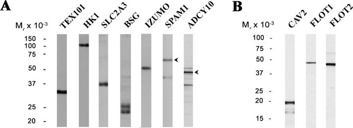
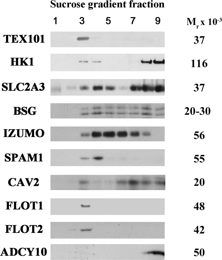
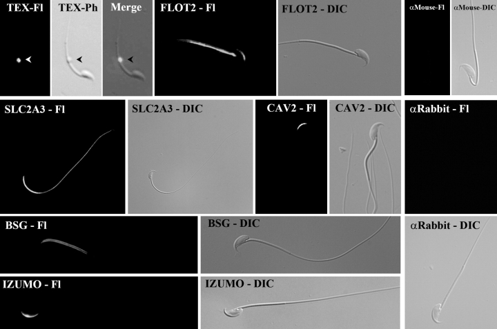
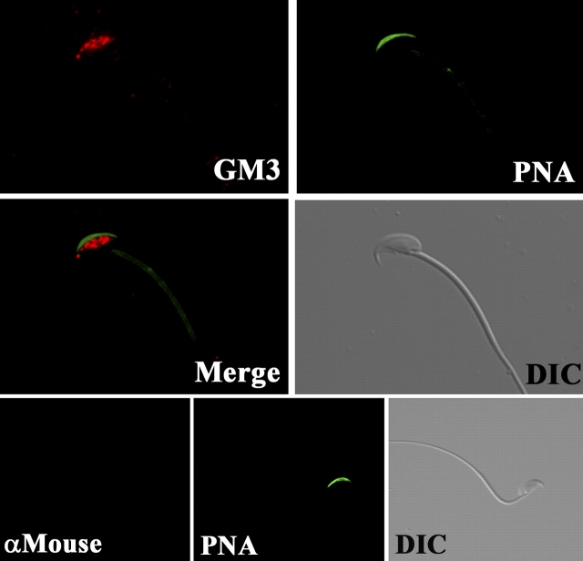
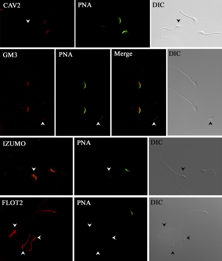
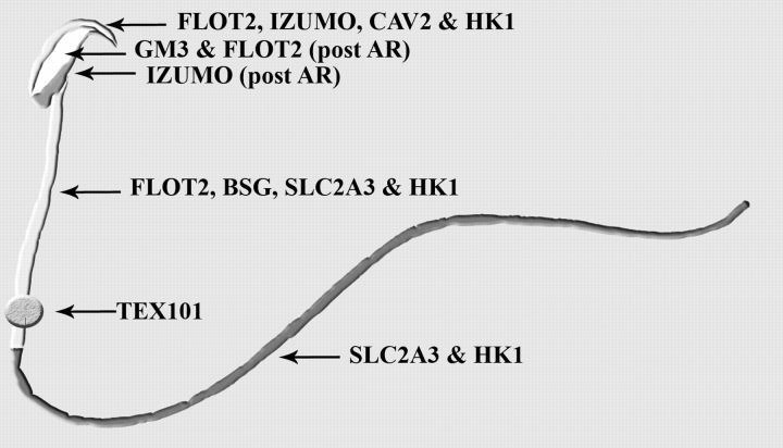
Mammalian sperm become fertile after completing capacitation, a process associated with cholesterol loss and changes in the biophysical properties of the sperm membranes that prepares the sperm to undergo the acrosome reaction. Different laboratories have hypothesized that cholesterol efflux can influence the extent and/or movement of lipid raft microdomains. In a previous study, our laboratory investigated the identity of sperm proteins putatively associated with rafts. After extraction with Triton X-100 and ultracentrifugation in sucrose gradients, proteins distributing to the light buoyant-density fractions were cored from polyacrylamide gels and microsequenced. In this study, a subset of these proteins (TEX101, basigin, hexokinase 1, facilitated glucose transporter 3, IZUMO, and SPAM1) and other molecules known to be enriched in membrane rafts (caveolin 2, flotillin 1, flotillin 2, and the ganglioside GM3) were selected to investigate their localization in the sperm and their behavior during capacitation and the acrosome reaction. These molecules localize to multiple sperm domains, including the acrosomal cap (IZUMO, caveolin 2, and flotillin 2), equatorial segment (GM3), cytoplasmic droplet (TEX101), midpiece (basigin, facilitated glucose transporter 3, and flotillin 2), and principal piece (facilitated glucose transporter 3). Some of these markers modified their immunofluorescence pattern after sperm incubation under capacitating conditions, and these changes correlated with the occurrence of the acrosome reaction. While GM3 and caveolin 2 were not detected after the acrosome reaction, flotillin 2 was found in the equatorial segment of acrosome-reacted sperm, and IZUMO distributed along the sperm head, reaching the post- and para-acrosomal areas. Taking into consideration the requirement of the acrosome reaction for sperm to become fusogenic, these results suggest that membrane raft dynamics may have a role in sperm-egg membrane interaction.


Figures

FIG. 1.
FIG. 1.
Specificity of antibodies directed toward DRMs and other sperm proteins. Total sperm extracts were prepared as detailed in Materials and Methods, separated using 10% (A) or 15% (B) acrylamide gels and analyzed by Western blot using different antibodies directed against the corresponding proteins. Arrowheads represent bands highlighted in Figure 2.
FIG. 2.
FIG. 2.
Distribution of proteins along the sucrose gradient. Sperm were extracted with 0.5% Triton X-100 and subjected to ultracentrifugation on a sucrose gradient as described in Materials and Methods. Nine fractions were collected from top to bottom and were analyzed by SDS-PAGE and Western blot using different antibodies. The molecular weights of the bands shown are in the right column. In the case of SPAM1 and ADCY10, bands shown are those marked with an arrowhead in Figure 1. A nonraft protein (ADCY10) was included as control.
FIG. 3.
FIG. 3.
Location of DRM proteins among the different sperm compartments. Mouse sperm were fixed with 2% formaldehyde, immobilized on slides, and permeabilized with 0.1% Triton X-100. Cells were incubated overnight with different antibodies raised in mouse (anti-TEX and anti-FLOT2) or rabbit (anti-SLC2A3, anti-CAV2, anti-BSG, and anti-IZUMO), followed by the respective Alexa Fluor-conjugated secondary antibody. Primary antibodies were omitted as control (αMouse and αRabbit panels). Illustrations represent the images obtained under fluorescence (Fl) or transmitted light illumination (Ph, phase contrast; DIC, differential interference contrast). Experiments were repeated at least three times; representative sperm are shown. Arrowheads represent cytoplasmic droplet. Original magnification ×60.
FIG. 4.
FIG. 4.
Detection of ganglioside GM3 in mouse sperm. Mouse sperm fixed and permeabilized as already described were incubated overnight with a monoclonal anti-GM3 antibody, followed by Alexa Fluor 555-conjugated secondary antibody (GM3). Cells were also stained with Alexa Fluor 488-conjugated PNA. No primary antibody controls were used (lower panels). Experiments were repeated at least three times; representative sperm are shown. DIC, differential interference contrast. Original magnification ×60.
FIG. 5.
FIG. 5.
Differential immunofluorescence pattern of GM3, CAV2, IZUMO, and FLOT2 in capacitated sperm. Mouse sperm incubated under capacitating conditions were analyzed by immunofluorescence as described in Materials and Methods using anti-GM3, anti-CAV2, anti-IZUMO, and anti-FLOT2, followed by the respective Alexa Fluor 555-conjugated secondary antibody. Cells were also stained with Alexa Fluor 488-conjugated PNA to relate the staining patterns to the acrosomal status. Sperm that underwent the acrosome reaction correspond to those showing the new staining patterns (arrowheads). Experiments were repeated at least three times; representative sperm are shown. DIC, differential interference contrast. Original magnification ×60.
FIG. 6.
FIG. 6.
Differential location of FLOT2 and IZUMO in intact and acrosome-reacted sperm. Mouse sperm were analyzed by immunofluorescence with anti-FLOT2 or anti-IZUMO before or after capacitation and treatment with the calcium ionophore A23187. Cells were also stained with Alexa Fluor 488-conjugated PNA to check for the acrosomal status. Experiments were repeated al least three times; representative sperm are shown. Original magnification ×60.
FIG. 7.
FIG. 7.
Schematic representation of the distribution of DRM proteins in sperm compartments depicting the results obtained using the antibodies directed against the different DRM-resident molecules analyzed in the study; HK1 localization as described previously [49] is also shown. As already noted in the text, IZUMO and FLOT2 demonstrate a different immunofluorescence pattern after the acrosome reaction (AR).

References

    1. Yanagimachi R.Mammalian fertilization. Knobil E, Neill JD.The Physiology of Reproduction, vol. 1, 2nd ed New York:Raven Press;1994: 189–317.
    1. Visconti PE, Kopf GS.Regulation of protein phosphorylation during sperm capacitation. Biol Reprod 1998; 59: 1–6. - PubMed
    1. Visconti PE, Bailey JL, Moore GD, Pan D, Olds-Clarke P, Kopf GS.Capacitation of mouse spermatozoa, I: correlation between the capacitation state and protein tyrosine phosphorylation. Development 1995; 121: 1129–1137. - PubMed
    1. Visconti PE, Moore GD, Bailey JL, Leclerc P, Connors SA, Pan D, Olds-Clarke P, Kopf GS.Capacitation of mouse spermatozoa, II: protein tyrosine phosphorylation and capacitation are regulated by a cAMP-dependent pathway. Development 1995; 121: 1139–1150. - PubMed
    1. Visconti PE, Ning X, Fornes MW, Alvarez JG, Stein P, Connors SA, Kopf GS.Cholesterol efflux-mediated signal transduction in mammalian sperm: cholesterol release signals an increase in protein tyrosine phosphorylation during mouse sperm capacitation. Dev Biol 1999; 214: 429–443. - PubMed

Publication types