. 2009 Jan 7;16(1):86-95.
doi: 10.1101/lm.1178309. Print 2009 Jan.

Interactions between prefrontal cortex and cerebellum revealed by trace eyelid conditioning

Affiliations

Interactions between prefrontal cortex and cerebellum revealed by trace eyelid conditioning

Brian E Kalmbach et al. Learn Mem. .

Abstract

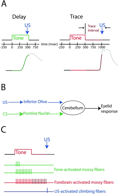
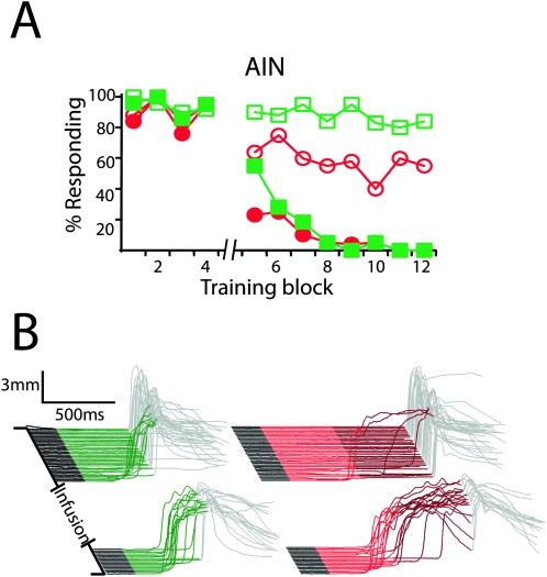
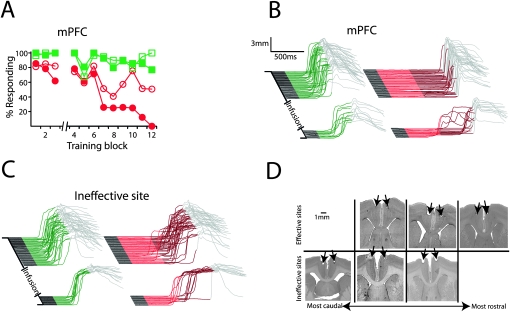
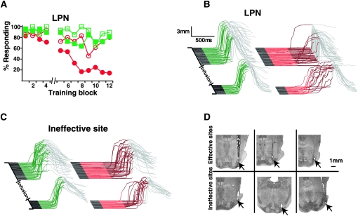
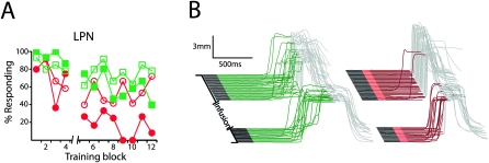
Eyelid conditioning has proven useful for analysis of learning and computation in the cerebellum. Two variants, delay and trace conditioning, differ only by the relative timing of the training stimuli. Despite the subtlety of this difference, trace eyelid conditioning is prevented by lesions of the cerebellum, hippocampus, or medial prefrontal cortex (mPFC), whereas delay eyelid conditioning is prevented by cerebellar lesions and is largely unaffected by forebrain lesions. Here we test whether these lesion results can be explained by two assertions: (1) Cerebellar learning requires temporal overlap between the mossy fiber inputs activated by the tone conditioned stimulus (CS) and the climbing fiber inputs activated by the reinforcing unconditioned stimulus (US), and therefore (2) trace conditioning requires activity that outlasts the presentation of the CS in a subset of mossy fibers separate from those activated directly by the CS. By use of electrical stimulation of mossy fibers as a CS, we show that cerebellar learning during trace eyelid conditioning requires an input that persists during the stimulus-free trace interval. By use of reversible inactivation experiments, we provide evidence that this input arises from the mPFC and arrives at the cerebellum via a previously unidentified site in the pontine nuclei. In light of previous PFC recordings in various species, we suggest that trace eyelid conditioning involves an interaction between the persistent activity of delay cells in mPFC-a putative mechanism of working memory-and motor learning in the cerebellum.


Figures

Figure 1.
Figure 1.
The procedures, neural pathways, and putative signals involved in delay and trace eyelid conditioning. (A) Stimulus timing for delay (left) and trace (right) training trials. For delay conditioning, the US overlaps in time with the tone CS. In this and subsequent figures, green is used to indicate the presentation of the CS for delay conditioning. For trace conditioning, the US is presented after CS offset, and “trace interval” refers to the period between CS offset and US onset. For convenience, we used red and maroon regions to represent the CS and trace interval, respectively. Sample conditioned eyelid responses are shown below, for which an upward deflection indicates closure of the eyelid. (B) Schematic representation of the pathways engaged by delay conditioning. The CS and US, respectively, engage mossy fibers and climbing fibers relatively directly, and forebrain input is not required for normal learning. (C) The signals hypothesized to engage the cerebellum during trace conditioning. The activity of mossy fibers directly activated by the tone CS does not significantly outlast the stimulus. Thus, a forebrain structure is thought to provide an input that overlaps in time with the US and is necessary to produce cerebellar learning.
Figure 2.
Figure 2.
The timing of inputs driven directly by trace conditioning stimuli in the absence of the forebrain is beyond the capabilities of cerebellar learning. (A) Direct electrical stimulation of mossy fibers as the CS failed to support trace conditioning with a 500-msec trace interval (red circles and squares; n = 9) but subsequently supported delay conditioning at 500 msec (green circles; n = 5) and 1000 msec (green squares; n = 4) ISIs. (B) Response traces of the last conditioning session of each protocol from representative animals. In this and subsequent figures, an upward deflection of the trace in the colored region represents a learned response and in the gray region represents a reflexive blink to the US. Trials in this and subsequent figures are stacked from first on the bottom to last on the top.
Figure 3.
Figure 3.
The ability of subjects to learn trace conditioning with a mossy fiber stimulation CS decreases as the trace interval increases. (A) Average response rates, normalized to delay conditioning trials, from the session after criterion was reached (n = 6). (B) Coronal section through the cerebellum shows a representative stimulation site in the middle cerebellar peduncle for the studies in this figure and in Figure 2. All stimulation sites were in the middle cerebellar peduncle. (C) Representative traces from the session after criterion was reached for trace intervals of 200, 300, and 400 msec.
Figure 4.
Figure 4.
Learning during dual delay/trace conditioning. (Left) The acquisition of delay responses (green squares) was faster than the acquisition of trace responses (red circles) during dual delay/trace training. Data points represent the mean response rate during nine-trial blocks of training (n = 11). There were 12 blocks per training session. Note that both delay and trace responses decrease during each session. We also observe this phenomenon in animals trained with only trace or only delay conditioning. This effect increases with the ISI and will be presented in a forthcoming paper along with a more detailed description and mechanistic implications. (Right) The faster acquisition of delay conditioning can also be seen in the trials needed to reach a criterion level of responding (see Materials and Methods). **P < 0.01.
Figure 5.
Figure 5.
The AIN is necessary for the expression of delay and trace conditioning. (A) Inactivating the AIN with muscimol after the fourth block of dual delay/trace conditioning (break in abscissa) abolished delay (filled green squares) and trace responses (filled red circles), while infusing ACSF had no effect (open squares indicate delay; open circles, trace; n = 5). (B) Representative traces taken from a muscimol infusion session. Delay and trace responses in this and subsequent figures have been separated for clarity but were intermixed during testing.
Figure 6.
Figure 6.
The mPFC is necessary for the expression of trace, but not delay, conditioning. (A) Infusing muscimol bilaterally into the mPFC after the third block of dual delay/trace conditioning (break in abscissa) abolished trace (filled red circles) but not delay responses (filled green squares), while infusing ACSF had no affect (open squares indicate delay; open circles, trace; n = 6). (B) Representative traces taken from an effective muscimol infusion session (same subject as shown in D, top left). (C) Representative traces taken from an ineffective muscimol infusion session (same subject as in D, bottom right). Note that in this example, neither trace nor delay responses were affected by the infusion. (D) Histological verification of cannula placements revealed that all cannula placements were in the vicinity of the caudal mPFC. Closer examination revealed that effective infusion sites tended to be more rostral and ventral than ineffective sites, suggesting that the necessary site(s) for trace eyelid conditioning is in the anterior cingulate and/or prelimbic cortices.
Figure 7.
Figure 7.
Mossy fibers originating in the LPN are necessary for the expression of trace, but not delay, conditioning. (A) Infusing muscimol into the LPN after the fourth block of dual delay/trace conditioning (break in abscissa) abolished trace (filled red circles) but not delay responses (filled green squares), while infusing ACSF had no affect (open squares indicate delay; open circles, trace; n = 8 for muscimol and 5 for ACSF). (B) Representative traces taken from an effective muscimol infusion session (same subjects as shown in D, top middle). (C) Representative traces taken from an ineffective muscimol infusion session (same subjects as in D, bottom right). (D) Histological verification of cannula placements revealed that effective infusion sites were located in the LPN, while ineffective sites were located near (<1mm), but outside of, the LPN.
Figure 8.
Figure 8.
The necessity of the LPN in trace conditioning is due to the trace interval and not a longer ISI. For these experiments, the trace conditioning tone was 100 msec, the trace interval was 500 msec, and the delay conditioning ISI was 1000 msec. (A) Infusing muscimol into the LPN after the fourth block of dual delay/trace conditioning (break in abscissa) abolished trace (filled red circles) but not delay responses (filled green squares), while infusing ACSF had no affect (open squares indicate delay; open circles, trace; n = 3). (B) Representative traces taken from an effective muscimol infusion session.

References

    1. Aitkin L.M., Boyd J. Acoustic input to the lateral pontine nuclei. Hear. Res. 1978;1:67–77. - PubMed
    1. Bao S., Chen L., Thompson R.F. Learning- and cerebellum-dependent neuronal activity in the lateral pontine nucleus. Behav. Neurosci. 2000;114:254–261. - PubMed
    1. Beylin A.V., Gandhi C.C., Wood G.E., Talk A.C., Matzel L.D., Shors T.J. The role of the hippocampus in trace conditioning: Temporal discontinuity or task difficulty? Neurobiol. Learn. Mem. 2001;76:447–461. - PubMed
    1. Bodner M., Kroger J., Fuster J.M. Auditory memory cells in dorsolateral prefrontal cortex. Neuroreport. 1996;7:1905–1908. - PubMed
    1. Boyd J., Aitkin L.M. Responses of single units in the pontine nuclei of the cat to acoustic stimulation. Neurosci. Lett. 1976;3:259–263. - PubMed

Publication types

MeSH terms

LinkOut - more resources