Review
. 2009 Feb;10(2):100-12.
doi: 10.1038/nrn2559. Epub 2009 Jan 15.

DNA repair deficiency and neurological disease

Affiliations
Review

DNA repair deficiency and neurological disease

Peter J McKinnon. Nat Rev Neurosci. 2009 Feb.

Erratum in

  • Nat Rev Neurosci. 2009 Mar;10(3):242

Abstract

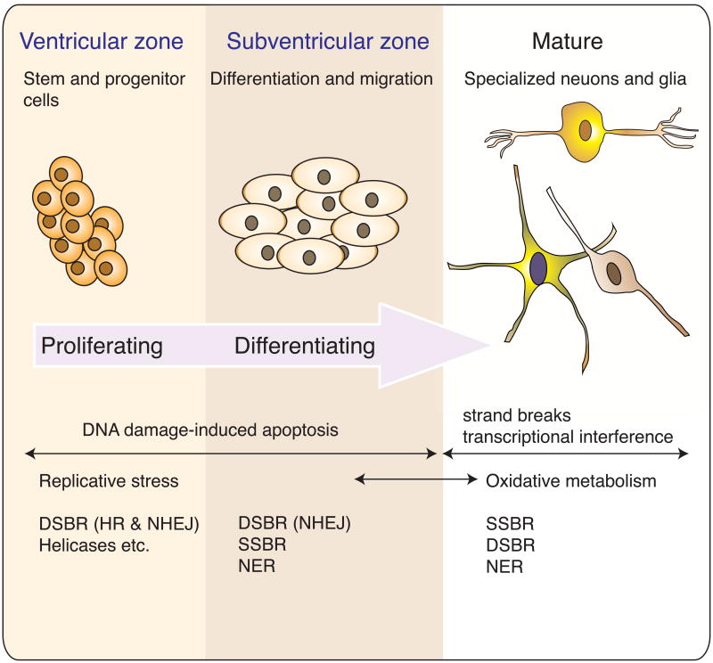
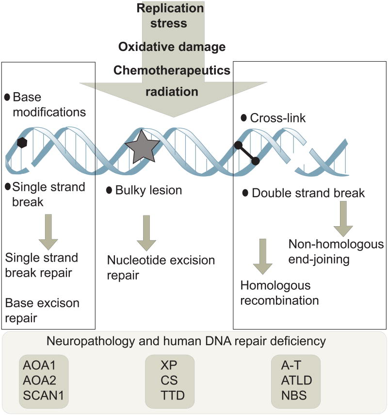
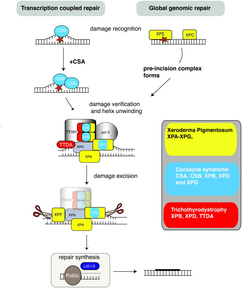
The ability to respond to genotoxic stress is a prerequisite for the successful development of the nervous system. Mutations in various DNA repair factors can lead to human diseases that are characterized by pronounced neuropathology. In many of these syndromes the neurological component is among the most deleterious aspects of the disease. The nervous system poses a particular challenge in terms of clinical intervention, as the neuropathology associated with these diseases often arises during nervous system development and can be fully penetrant by childhood. Understanding how DNA repair deficiency affects the nervous system will provide a rational basis for therapies targeted at ameliorating the neurological problems in these syndromes.


Figures

Figure 1
Figure 1. DNA damage and repair during nervous system development
During development the formation of the nervous system occurs via widespread proliferation, migration and differentiation. The diversity of the nervous system begins as stem/progenitors cells that divide in the ventricular zone and also, albeit to a lesser extent, the subventricular zone and then undergo differentiation, migration and maturation to give rise to the neurons and glia of the adult nervous system. A wide variety of functionally specialized cells with unique properties all originate from proliferative cells in one of four ventricles present in the nervous system. At different stages of development the nervous system is susceptible to different types of DNA damage. During proliferation, replication associated DNA strand breaks can occur that may require DNA double strand break repair (DSBR), involving homologous recombination (HR) or non-homologous end-joining (NHEJ) and associated functions of helicases and various other replication components that interface with DNA repair. In differentiating cells repair options are more limited as HR is not available after the cells exit the cell cycle. At this stage, NHEJ repairs DNA DSBs while other types of DNA damage require nucleotide excision repair (NER) or single strand break repair (SSBR). Because the nervous system can easily replace cells during development, DNA damage-induced apoptosis is also a frequent outcome at these stages of neural development. In the mature nervous system, strand breaks and DNA modification from oxidative damage engage SSBR, NER and DSBR and DNA damage that is not repaired can disrupt transcription leading to cell death.
Figure 2
Figure 2. Types of DNA damage and repair
A variety of different types of DNA damage can occur in neural cells as a result of endogenous agents such as replication stress or free radicals from oxidative metabolism. Exogenous insults such as ionizing or ultraviolet radiation and chemotherapeutics can also cause different types of DNA damage. These agents can cause single or double strand breaks in the DNA, base modifications, helix-distorting bulky lesions or cross-links of DNA strands. Biochemically distinct DNA repair pathways are available to repair each class of DNA damage. DNA repair pathways that are particularly important for nervous system function comprise pathways that repair DNA single and double strand breaks and nucleotide excision repair. When any of these pathways are defective, diseases can result that impact the nervous system; representative examples of human syndromes linked to defects in the particular DNA repair pathways are listed. Defective repair of DNA single strand breaks can lead to ataxia with oculomotor apraxia 1 or 2 (AOA1 and AOA2) or spinocerebellar ataxia with axonal neuropathy 1 (SCAN1), while defects in nucleotide excision repair can lead to xeroderma pigmentosum (XP), Cockayne Syndrome (CS) or trichothyrodystrophy (TTD). Defective responses to DNA double strand breaks can lead to ataxia telangiectasia (A-T), A-T like disease (ATLD) or Nijmegen breakage syndrome (NBS).
Figure 3
Figure 3. Repairing DNA strand breaks
The repair of double strand breaks and single strand breaks occur by distinct biochemical pathways. Two different repair pathways can deal with a DSB, and the choice depends on the proliferative state of the cell. A. Homologous recombination can be utilized in proliferating cells. Replication Protein A (RPA) coats the resected single stranded DNA and recruits RAD51 recombinase, which together with a multitude of other factors, , , including BRCA2 and XRCC2, , repairs the DNA via a processes whereby template DNA from the sister-chromatid is inserted into the damaged chromatid and acts as an error-free template. Ligation of the break requires DNA ligase I (LIG1). B. Non-homologous end-joining (NHEJ) can function in proliferating and non-proliferating cells. Heterodimeric KU70 and KU80 bind DNA ends and recruit DNA-PK (catalytic subunit of the DNA-dependent kinase) which, together with XRCC4, XLF and DNA ligase IV (LIG4), reseals the DNA ends after suitable end processing from various nucleases-. Because some nucleotides may be lost in this process, NHEJ is often referred to as error-prone repair. However, DNA breaks are repaired, and so these ‘errors’ may generally be of little consequence in non-dividing cells like neurons. C. Strand breaks can arise from damaged bases being removed via specific DNA glycosylases (referred to as base excision repair) or direct backbone damage that severs the DNA strand. In both cases the stand break leads to Poly ADP-Ribose Polymerase (PARP) accumulation, which facilitates recruitment of the XRCC1 scaffolding protein that is important for promoting repair. XRCC1 recruits repair factors that modify the DNA ends for re-ligation. Depending upon the nature of the DNA ends present at the SSB, TDP1 (Tyrosyl DNA Phosphodiesterase 1; 3′ modified ends or topoisomerase I DNA adducts) or APTX (Aprataxin; 5′ends resulting from adenylated DNA from abortive ligation events) may be required to modify the damaged DNA for repair. Repair can involve removal of a single nucleotide (short patch repair) or a longer patch of nucleotide (long patch repair) by PCNA and Ligase 1.
Figure 4
Figure 4. ATM signalling in response to DNA damage
ATM (Ataxia-Telangiectasia, Mutated) is a serine/threonine protein kinase modulated by the MRN (Mre11-Rad50-NBS1) complex that is critically important for responding to DNA DSBs, and loss of these factors can lead to profound neuropathology. Many ATM substrates are important cell cycle and apoptotic regulators. A primary function of ATM after DNA damage is checkpoint activation. At each phase of the cell cycle ATM activates checkpoint proteins, amongst which are FANCD2 (Fanconi Anemia group D2), SMC1 (Structural Maintenance of Chromosomes 1), NBS1 (Nijmegen Breakage Syndrome 1), CDC25C (cell division cycle 25C) and BRCA1 (Breast Cancer Associated 1); an inclusive list of ATM substrates is available, . Key ATM substrates are p53 and Chk2 (checkpoint kinase 2), which are responsible for activation of the G1 checkpoint and apoptosis. A main function of ATM in the nervous system may be to modulate DNA damage induced apoptosis. This is because in the nervous system, the prolific cell division that occurs allows damaged cells to be easily replaced, and so ATM fulfils an important genome monitoring function as immature cells exit the cell cycle, whereby cells with DNA damage are eliminated in an ATM dependent manner.
Figure 5
Figure 5. Nucleotide excision repair and related diseases
Defects in nucleotide excision repair (NER) lead to at least three human syndromes characterized by neurodegeneration and DNA repair; xeroderma pigmentosum, Cockayne syndrome and Trichothiodystrophy (also see Table 1). NER can involve transcription-coupled repair (TCR), which occurs when the transcription complex encounters a damaged template and the more general repair pathway, global genomic repair (GGR), which repairs lesions present in non-transcribed DNA. The difference between these pathways is at the step of damage detection. The sequence of events in NER involves DNA damage recognition followed by an incision, damage removal and repair synthesis. DNA damage recognition in TCR involves initiation by CSB after the RNA Polymerase II complex encounters damage. In GGR damage is recognized by XPE or XPC. Pre-incision events involve XPA/RPA. The TFIIH complex, including the XPB and XPD helicases, unwinds the DNA helix resulting in an open conformation. XPG is then recruited to the complex and binds to TFIIH and RPA. The Cockayne syndrome proteins, CSA and CSB, are also involved in TCR and may function in shifting TFIIH from a transcription to repair complex. Dual incision involves XPG performing the initial cleavage 3′ to the site of DNA damage, followed by 5′ cleavage through the action or the XPF nuclease. Repair synthesis is performed by DNA Polymerase δ or ε with PCNA, and ligation to seal the gap involves DNA ligase I. Three main classes of diseases resulting from disruption of NER are listed, and pathway components that are disrupted in the respective diseases are represented in corresponding colors.

References

    1. Bender A, et al. High levels of mitochondrial DNA deletions in substantia nigra neurons in aging and Parkinson disease. Nat Genet. 2006;38:515–7. - PubMed
    1. Katyal S, McKinnon PJ. DNA strand breaks, neurodegeneration and aging in the brain. Mech Ageing Dev. 2008;129:483–91. - PMC - PubMed
    1. Kraytsberg Y, et al. Mitochondrial DNA deletions are abundant and cause functional impairment in aged human substantia nigra neurons. Nat Genet. 2006;38:518–20. - PubMed
    1. Nouspikel T, Hanawalt PC. When parsimony backfires: neglecting DNA repair may doom neurons in Alzheimer's disease. Bioessays. 2003;25:168–73. - PubMed
    1. Chan WY, Lorke DE, Tiu SC, Yew DT. Proliferation and apoptosis in the developing human neocortex. Anat Rec. 2002;267:261–76. - PubMed

Publication types

MeSH terms