. 2009;4(1):e4204.
doi: 10.1371/journal.pone.0004204. Epub 2009 Jan 15.

Respiratory dendritic cell subsets differ in their capacity to support the induction of virus-specific cytotoxic CD8+ T cell responses

Affiliations

Respiratory dendritic cell subsets differ in their capacity to support the induction of virus-specific cytotoxic CD8+ T cell responses

Taeg S Kim et al. PLoS One. 2009.

Abstract

Dendritic cells located at the body surfaces, e.g. skin, respiratory and gastrointestinal tract, play an essential role in the induction of adaptive immune responses to pathogens and inert antigens present at these surfaces. In the respiratory tract, multiple subsets of dendritic cells (RDC) have been identified in both the normal and inflamed lungs. While the importance of RDC in antigen transport from the inflamed or infected respiratory tract to the lymph nodes draining this site is well recognized, the contribution of individual RDC subsets to this process and the precise role of migrant RDC within the lymph nodes in antigen presentation to T cells is not clear. In this report, we demonstrate that two distinct subsets of migrant RDC--exhibiting the CD103(+) and CD11b(hi) phenotype, respectively--are the primary DC presenting antigen to naïve CD4(+) and CD8(+) T lymphocytes in the draining nodes in response to respiratory influenza virus infection. Furthermore, the migrant CD103(+) RDC subset preferentially drives efficient proliferation and differentiation of naive CD8(+) T cells responding to infection into effector cells, and only the CD103(+) RDC subset can present to naïve CD8(+) T cells non-infectious viral vaccine introduced into the respiratory tract. These results identify CD103(+) and CD11b(hi) RDC as critical regulators of the adaptive immune response to respiratory tract infection and potential targets in the design of mucosal vaccines.


Conflict of interest statement

Competing Interests: The authors have declared that no competing interests exist.

Figures

Figure 1
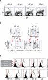
Figure 1. DC subsets in the normal and influenza-infected lung.
(A and C) Conventional RDC subsets in the normal and influenza-infected lung. Hematopoietic origin cells in the lung suspensions are identified as CD45+. Among the CD45+CD11c+ cells, the highly auto-fluorescent alveolar macrophages (AM) and B220+ plasmacytoid DC are excluded from further analyses (see Fig. S1 A and B for gating and identification strategy). CD103+ RDC (f), MoRDC (g) and CD11bhi RDC (h) in the uninfected (left) or infected (at d2 p.i., right) lung are identified (A) and enumerated (C) at indicated days p.i. (B) Ability of RDC subsets to take up soluble antigens in vivo. Mice are given i.n. FITC-labeled ovalbumin (FITC-Ova) or unlabeled Ova proteins 1 hr prior to sacrifice. The amounts (%) of FITC-Ova taken up by RDC subsets are compared to those of ova, and the numbers in the inserts indicate mean fluorescent intensity (MFI) of FITC-Ova (n>3).
Figure 2
Figure 2. Progressive accumulation of CD103+ DC and CD11bhi DC in the lung-draining lymph nodes of influenza-infected mice.
(A) Accumulation of CD11chiMHC IIhi DC in the MLN after influenza infection. Representatives of total CD11c+ cells in the MLN of influenza-infected mice at indicated days p.i. are shown. (B) Accumulation of three major DC subsets in the MLN of influenza-infected mice. CD103+ DC (CD11chiMHC IIhiCD103+CD11b+/−, group I), CD11bhi DC (CD11chiMHC IIhiCD103CD11bhi, group II) and Gr-1+ MoDC (CD11cloMHC IIloCD103Gr-1+CD11bhi, group III) are highly represented in the MLN at d3 p.i. (right panel) compared to those in uninfected animals (left panel) (see gating strategy in Fig. S2). (C) Phenotypic features of DC subsets in the MLN of influenza-infected mice at d3 p.i. Surface marker expression on CD103+ DC (Group I, gray), CD11bhi DC (Group II, white) and Gr-1+ MoDC (Group III, black) identified in the MLN of infected (d3 p.i.) mice (right panel in B) is examined after staining with indicated mAbs. Vertical lines (|) indicate the median values of surface staining with isotype-matched control antibodies.
Figure 3
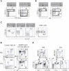
Figure 3. Sources of DC subsets in the MLN of influenza-infected mice.
(A and B) Acquisition and transportation of FITC-Ova (A) or viral proteins (B) by lung-residing DC into the MLN of infected mice. (A) Fluorescent (FITC)-tagged or unlabeled Ova proteins were i.n. instilled into influenza-infected mice at d2 p.i. One day later, DC subsets in the MLN of the Ova-instilled infected mice are examined for the uptake (%) of FITC-Ova. (B) The specific % NP+ DC subsets in the MLN of infected mice at d3 p.i. is examined after intracellular staining for viral nucleoprotein (NP). (C) Examination of local proliferation of DC subsets in the MLN of infected mice. DC isolated from the MLN of i.n. infected mice at d3 p.i. are intracellularly stained for an nuclear antigen Ki-67. Ki-67 staining on CD8+ T cells isolated from MLN of infected mice at d7 p.i. are depicted as a positive control. (D and E) Accumulation of CD11bhi DC and CD103+ DC in the MLN of infected mice requires the presence of the corresponding counterparts in the lung. (D) Naïve DTR mice are instilled i.n. with DTx (lower panel) or vehicle alone (top panel), and depletion of the CD11c+ RDC subsets in the lung is examined 48 hr later (n>5). (E) DC subsets in the MLN of influenza-infected mice are identified at d3 p.i. in the DTR mice i.n. given either DTx or PBS one day prior to infection (n>10).
Figure 4
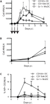
Figure 4. Kinetics of the accumulation of three major DC subsets in the MLN of influenza-infected mice.
(A and B) Mice were i.n. infected with influenza, and at various days p.i., Gr-1+ MoDC (□), CD11bhi DC (⋄) and CD103+ DC (xˆ) in the MLN of the infected mice are identified (as shown in Fig. S2) and enumerated (A). The magnitude of DC subsets accumulated in the MLN from d0–2 p.i. in (A) is expanded in (B) (mean±SD; n = 4–8 mice/group). (C) Kinetics of the accumulation of NP+ DC subsets in the MLN of influenza-infected mice from d1 to d5 p.i. The % NP+ DC subsets – Gr-1+ MoDC (□), CD11bhi DC (⋄), and CD103+ DC (xˆ) – are represented after subtracting that of non-specific staining with an irrelevant mAb (mean±SD; n = 4–5 mice/group).
Figure 5
Figure 5. The CD11bhi and CD103+ RDC subsets are required for the induction of optimal proliferative expansion of virus-specific CD8+ T cells responding to respiratory influenza virus infection in vivo.
(A–D) Ablation of both lung-residing CD11bhi and CD103+ DC subsets prior to infection resulted in a near absence of virus-specific naïve CL-4 CD8+ T cell proliferation in response to respiratory (A and B), but not intravenous (C and D), influenza virus infection. Following CFSE-labeled CL-4 CD8+ T cell transfer into DTR mice, DTx (or PBS) is administered i.n. one day prior to infection. Four (i.n.) or 3 (i.v.) days after virus infection, the division profiles of CL-4 T cells (A or C) and total recovery of the number of CL-4 T cells (left in B or D) and total cellularity (right in B or D) in the MLN of infected DTR mice were determined (mean±SD; n = 4–6).
Figure 6
Figure 6. Airway-derived migrant CD103+ DC in the inflamed MLN are superior in presenting or cross-presenting influenza virus antigens to naïve CD8+ T cells.
(A–C) Efficiency of DC subsets in the MLN of infected animals to stimulate naïve either CD8+ or CD4+ T cells after intranasal infection with infectious influenza virus. DC subsets isolated from the MLN of mice infected 3 d earlier with influenza virus were co-cultured with CFSE-labeled HA-specific naïve TCR tg CD8+ or CD4+ T cells for 4 days. Proliferation profile (A) or total recovery (B) of viable CD8+ or CD4+ T cells stimulated by the MLN-derived DC subsets are shown (mean±SD; n = 5). Effector functions of CD8+ T lymphocytes stimulated by either CD11bhi DC or CD103+ DC are examined at d4 co-culture (C). (D) Efficiency of DC subsets in the MLN to stimulate naïve CD8+ or CD4+ T cells after intranasal administration of non-infectious influenza virions. DC subsets isolated from the MLN of mice inoculated 3 d earlier with inactivated influenza virus were co-cultured with CFSE-labeled HA-specific naïve TCR tg CD8+ or CD4+ T cells for 4 days. Representatives of proliferation profile of T cell subsets are depicted (n = 3).

Similar articles

Cited by

References

    1. Banchereau J, Steinman RM. Dendritic cells and the control of immunity. Nature. 1998;392:245–252. - PubMed
    1. Henri S, Vremec D, Kamath A, Waithman J, Williams S, et al. The dendritic cell populations of mouse lymph nodes. J Immunol. 2001;167:741–748. - PubMed
    1. Villadangos JA, Heath WR. Life cycle, migration and antigen presenting functions of spleen and lymph node dendritic cells: limitations of the Langerhans cells paradigm. Semin Immunol. 2005;17:262–272. - PubMed
    1. Stumbles PA, Upham JW, Holt PG. Airway dendritic cells: co-ordinators of immunological homeostasis and immunity in the respiratory tract. Apmis. 2003;111:741–755. - PubMed
    1. Forster R, Schubel A, Breitfeld D, Kremmer E, Renner-Muller I, et al. CCR7 coordinates the primary immune response by establishing functional microenvironments in secondary lymphoid organs. Cell. 1999;99:23–33. - PubMed