

Hematopoietic- and neurologic-expressed sequence 1 expression in the murine GL261 and high-grade human gliomas
- PMID: 19145478
- PMCID: PMC2819207
- DOI: 10.1007/s12253-008-9147-4
Hematopoietic- and neurologic-expressed sequence 1 expression in the murine GL261 and high-grade human gliomas
Abstract
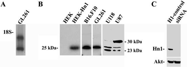
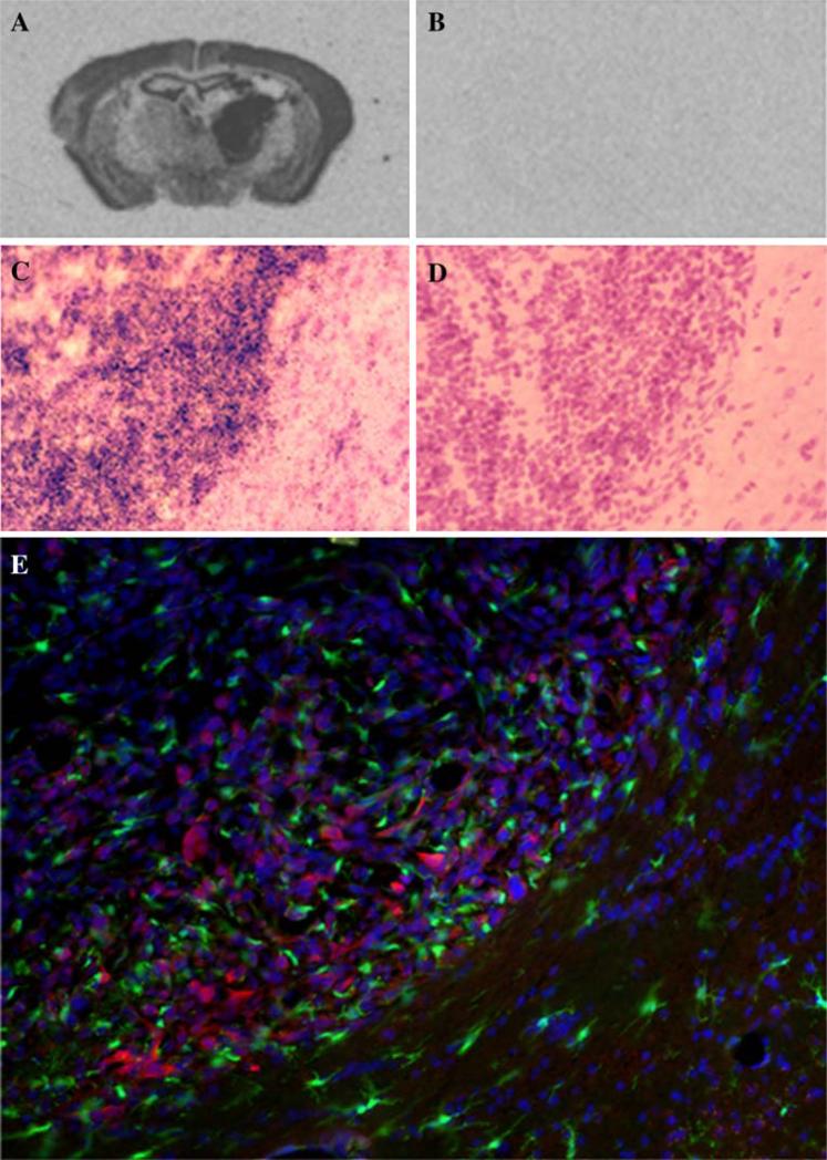
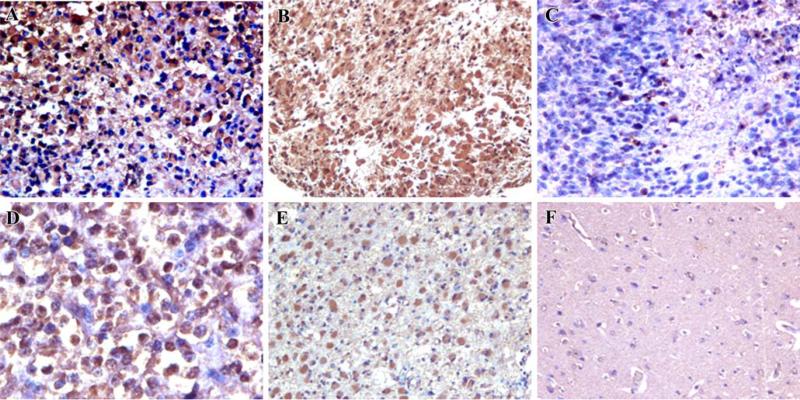
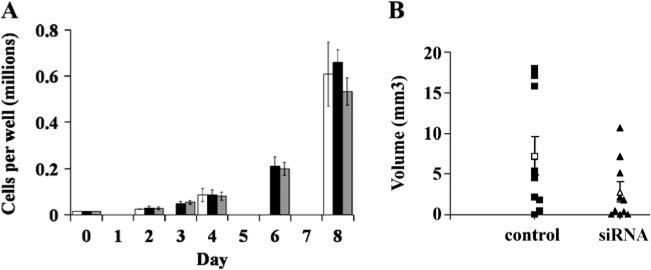
The hematopoietic- and neurologic-expressed sequence 1 (Hn1) gene encodes a highly conserved protein that is expressed in developing and regenerating tissues. In this study, Hn1 expression was evaluated in human and murine malignant gliomas. Hn1 mRNA and protein were detected in the murine GL261 glioma cell line and in GL261 brain tumors in vivo. HN1 is also expressed in human U118MG and U87MG cell lines. Evaluation of human brain tumors using an anti-Hn1 polyclonal antibody detected strong immunoreactivity in high-grade (WHO III and IV) malignant gliomas. The rate of GL261 cell proliferation in vitro was unaltered by Hn1 depletion using an anti-Hn1 siRNA. However, tumors established from Hn1-depleted GL261 cells formed significantly smaller volumes than those established from control-treated cells. These data suggest a role for Hn1 in the biology of malignant brain tumors.
Figures
Similar articles
-
Hematopoietic- and neurologic-expressed sequence 1 (Hn1) depletion in B16.F10 melanoma cells promotes a differentiated phenotype that includes increased melanogenesis and cell cycle arrest.Differentiation. 2009 Jul;78(1):35-44. doi: 10.1016/j.diff.2009.04.001. Epub 2009 May 7. Differentiation. 2009. PMID: 19427096 Free PMC article.
-
Differential expression of p42.3 in low- and high-grade gliomas.World J Surg Oncol. 2014 Jun 14;12:185. doi: 10.1186/1477-7819-12-185. World J Surg Oncol. 2014. PMID: 24927751 Free PMC article.
-
Overexpression of septin 7 suppresses glioma cell growth.J Neurooncol. 2010 Jul;98(3):329-40. doi: 10.1007/s11060-009-0092-1. Epub 2009 Dec 25. J Neurooncol. 2010. PMID: 20035367
-
The Zfx gene is expressed in human gliomas and is important in the proliferation and apoptosis of the human malignant glioma cell line U251.J Exp Clin Cancer Res. 2011 Dec 20;30(1):114. doi: 10.1186/1756-9966-30-114. J Exp Clin Cancer Res. 2011. PMID: 22185393 Free PMC article.
-
[CDK1 expression and effects of CDK1 silencing on the malignant phenotype of glioma cells].Zhonghua Zhong Liu Za Zhi. 2007 Jul;29(7):484-8. Zhonghua Zhong Liu Za Zhi. 2007. PMID: 18069625 Chinese.
Cited by
-
HN1 contributes to migration, invasion, and tumorigenesis of breast cancer by enhancing MYC activity.Mol Cancer. 2017 May 11;16(1):90. doi: 10.1186/s12943-017-0656-1. Mol Cancer. 2017. PMID: 28490334 Free PMC article.
-
HN1L-mediated transcriptional axis AP-2γ/METTL13/TCF3-ZEB1 drives tumor growth and metastasis in hepatocellular carcinoma.Cell Death Differ. 2019 Nov;26(11):2268-2283. doi: 10.1038/s41418-019-0301-1. Epub 2019 Feb 18. Cell Death Differ. 2019. PMID: 30778199 Free PMC article.
-
HN1 as a diagnostic and prognostic biomarker for liver cancer.Biosci Rep. 2020 Jul 31;40(7):BSR20200316. doi: 10.1042/BSR20200316. Biosci Rep. 2020. PMID: 32700728 Free PMC article.
-
Increased expression of hematological and neurological expressed 1 (HN1) is associated with a poor prognosis of hepatocellular carcinoma and its knockdown inhibits cell growth and migration partly by down-regulation of c-Met.Kaohsiung J Med Sci. 2020 Mar;36(3):196-205. doi: 10.1002/kjm2.12156. Epub 2019 Nov 20. Kaohsiung J Med Sci. 2020. PMID: 31749294 Free PMC article.
-
Overexpression of HN1L promotes cell malignant proliferation in non-small cell lung cancer.Cancer Biol Ther. 2017 Nov 2;18(11):904-915. doi: 10.1080/15384047.2017.1385678. Epub 2017 Oct 20. Cancer Biol Ther. 2017. PMID: 29053395 Free PMC article.
References
-
- Zujovic V, Luo D, Baker HV, Lopez MC, Miller KR, Streit WJ, Harrison JK. The facial motor nucleus transcriptional program in response to peripheral nerve injury identifies Hn1 as a regeneration-associated gene. J Neurosci Res. 2005;82:581–591. - PubMed
-
- Goto T, Hisatomi O, Kotoura M, Tokunaga F. Induced expression of hematopoietic- and neurologic-expressed sequence 1 in retinal pigment epithelial cells during newt retina regeneration. Exp Eye Res. 2006;83:972–980. - PubMed
-
- Lu KH, Patterson AP, Wang L, Marquez RT, Atkinson EN, Baggerly KA, Ramoth LR, Rosen DG, Liu J, Hellstrom I, Smith D, Hartmann L, Fishman D, Berchuck A, Schmandt R, Whitaker R, Gershenson DM, Mills GB, Bast RC., Jr. Selection of potential markers for epithelial ovarian cancer with gene expression arrays and recursive descent partition analysis. Clin Cancer Res. 2004;10:3291–3300. - PubMed
-
- Tang W, Lai YH, Han XD, Wong PM, Peters LL, Chui DH. Murine Hn1 on chromosome 11 is expressed in hemopoietic and brain tissues. Mamm Genome. 1997;8:695–696. - PubMed
-
- Zhou G, Wang J, Zhang Y, Zhong C, Ni J, Wang L, Guo J, Zhang K, Yu L, Zhao S. Cloning, expression and subcellular localization of HN1 and HN1L genes, as well as characterization of their orthologs, defining an evolutionarily conserved gene family. Gene. 2004;331:115–123. - PubMed
Publication types
MeSH terms
Substances
Grants and funding
LinkOut - more resources
Full Text Sources
Medical
Molecular Biology Databases