Review
. 2008 Dec 18;8(14):20.1-21.
doi: 10.1167/8.14.20.

The significance of microsaccades for vision and oculomotor control

Affiliations
Review

The significance of microsaccades for vision and oculomotor control

Han Collewijn et al. J Vis. .

Abstract

Over the past decade several research groups have taken a renewed interest in the special role of a type of small eye movement, called 'microsaccades', in various visual processes, such as the activation of neurons in the central nervous system, or the prevention of image fading. As the study of microsaccades and their relation to visual processes goes back at least half a century, it seems appropriate to review the more recent reports in light of the history of research on maintained oculomotor fixation, in general, and on microsaccades in particular. Our review shows that there is no compelling evidence to support the view that microsaccades (or, fixation saccades more generally) serve a necessary role in improving oculomotor control or in keeping the visual world visible. The role of the retinal transients produced by small saccades during fixation needs to be evaluated in the context of both the brisk image motions present during active visual tasks performed by freely moving people, as well as the role of selective attention in modulating the strength of signals throughout the visual field.


Figures

Figure 1
Figure 1
Horizontal and vertical eye movements over time, as measured with the contact lens optical lever, while fixating a small point target showing a stable line of sight maintained either with (bottom traces) or without (top traces) the occurrence of microsaccades (from Steinman et al., 1973, Science, 181, 810–819). Reproduced with permission.
Figure 2
Figure 2
Distribution of sizes of microsaccades of 4 fixating human subjects, recorded with the contact lens technique (taken from Boyce, 1967, Proceedings of the Royal Society of London B, 167, 293–315). Reproduced with permission.
Figure 3
Figure 3
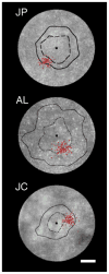
Area of highest cone density is not always used for fixation. Shown are retinal montages of the foveal cone mosaic for three subjects. The black square represents the foveal center of each subject. The dashed black line is the isodensity contour line representing a 5% increase in cone spacing, and the solid black line is the isodensity contour line representing a 15% increase in cone spacing. Red dots are individual fixation locations. Scale bar is 50 μm (from Putnam et al., 2005).
Figure 4
Figure 4
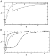
The effects of small, imposed target motion on a stabilized (foveal) image in restoring visibility. Figures taken from Ditchburn and Drysdale (1977b), Proceedings of the Royal Society of London B, 167, 385–406. Reproduced with permission. The Y-axis shows the level of visibility (Vc) to the subject; the X-axis shows the (peak-to-peak) magnitude (in min arc) of the imposed movement. Upper panel: effect of sinusoidal motion at 0.55 Hz (graph b refers to previous measurements by Ditchburn, Fender, & Mayne). Lower panel: effect of square wave motion (0.5 Hz. Graphs a and b refer to sharp targets; graph c to a low contrast target.
Figure 5
Figure 5
(A) Movements of eye and head during fixation while the head was either supported by a bite-board or was free to move while the subject was either sitting or standing as still as possible. Repetitive vertical stripes are 1-s time markers (from Skavenski et al., 1979). (B) Horizontal eye movements of one subject fixating a distant target while freely moving the head over small amplitudes. The head position trace shows head position scaled to 1/10 of its value. (C) Distribution of retinal image velocities (right eye) during small active head motions, as in (B). (B, C from Steinman & Collewijn, 1980, Vision Research, 20, 415–429). Reproduced with permission.
Figure 6
Figure 6
The relative distribution (%) of the sizes of all saccades made during a natural task (sequential fixation or finger-tapping of real physical targets in which subjects could freely move their heads and arm. Figures taken from Malinov et al. (2000), Vision Research, 40, 2083–2090. Reproduced with permission; all saccades from 4 subjects pooled. (A) Overall size distribution for all saccades (horizontal and vertical components shown each for the two tasks). (B) Distribution of size for all saccades smaller than 5 deg. (C) Distribution of size (2-D vector) of size for all saccades smaller than 1 deg. Total number of saccades is given each panel. Notice the virtual absence of real microsaccades (<12 min arc). (Instrument noise is constant at bit-noise of ±1 min arc).
Figure 7
Figure 7
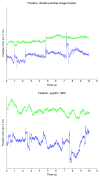
Sample records of horizontal and vertical eye movements taken from the same subject during fixation of a small stationary cross. Top records were made using a Dual Purkinje Image Tracker while the subject’s head was stabilized by a bite-board. Bottom records were made with an Eyelink 1000 mounted on a table while the head was stabilized by a chin and forehead rest. Green traces (top traces of each graph) show vertical movements, blue (bottom traces of each) show horizontal (Kowler, unpublished recordings).
Figure 8
Figure 8
Sample recordings of eye movements during fixation (from Møller et al., 2002, Graefe’s Archive for Clinical and Experimental Ophthalmology, 240, 765–770). Reproduced with permission.

References

    1. Arend LE, Timberlake GT. What is psychophysically perfect image stabilization? Do perfectly stabilized images always disappear? Journal of the Optical Society of America A, Optics and Image Science. 1986;3:235–241. - PubMed
    1. Bahcall DO, Kowler E. Attentional interference at small spatial separations. Vision Research. 1999;39:71–86. - PubMed
    1. Bahill AT, Adler D, Stark L. Most naturally occurring human saccades have magnitudes of 15 degrees or less. Investigative Ophthalmology. 1975;14:468–469. - PubMed
    1. Bahill AT, Kallman JS, Lieberman JE. Frequency limitations of the two-point central difference differentiation algorithm. Biological Cybernetics. 1982;45:1–4. - PubMed
    1. Ballard DH, Hayhoe MM, Pelz JB. Memory representations in natural tasks. Journal of Cognitive Neuroscience. 1995;7:66–80. - PubMed