Comparative Study
. 2009 Feb 10;48(5):1077-83.
doi: 10.1021/bi801871h.

Evidence that alpha-synuclein does not inhibit phospholipase D

Affiliations
Comparative Study

Evidence that alpha-synuclein does not inhibit phospholipase D

Irit Rappley et al. Biochemistry. .

Abstract

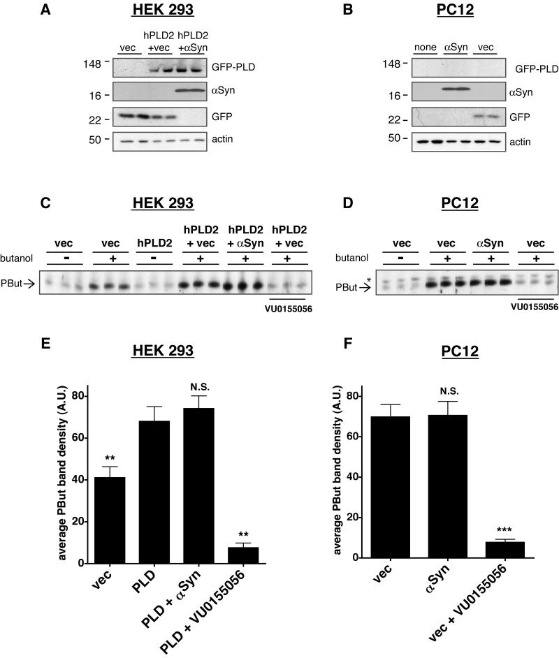
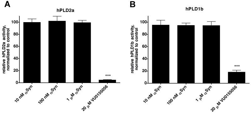
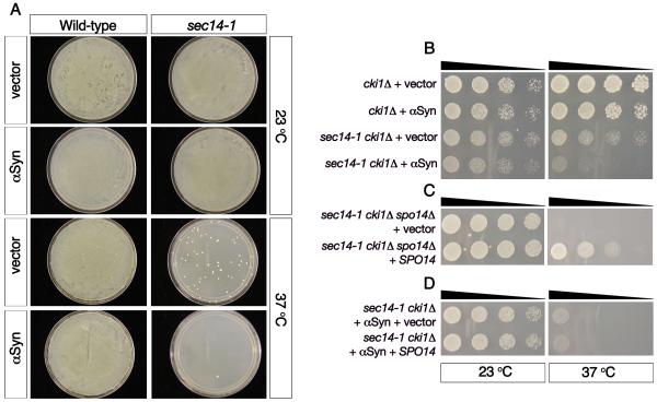
Alpha-synuclein (alphaSyn) is a small cytosolic protein of unknown function, which is highly enriched in the brain. It is genetically linked to Parkinson's disease (PD) in that missense mutations or multiplication of the gene encoding alphaSyn causes early onset familial PD. Furthermore, the neuropathological hallmarks of both sporadic and familial PD, Lewy bodies and Lewy neurites, contain insoluble aggregates of alphaSyn. Several studies have reported evidence that alphaSyn can inhibit phospholipase D (PLD), which hydrolyzes phosphatidylcholine to form phosphatidic acid and choline. Although various hypotheses exist regarding the roles of alphaSyn in health and disease, no other specific biochemical function for this protein has been reported to date. Because PLD inhibition could represent an important function of alphaSyn, we sought to extend existing reports on this interaction. Using purified proteins, we tested the ability of alphaSyn to inhibit PLD activity in cell-free assays. We also examined several cell lines and transfection conditions to assess whether alphaSyn inhibits endogenous or overexpressed PLD in cultured mammalian cells. In yeast, we extended our previous report of an interaction between alphaSyn and PLD-dependent phenotypes, for which PLD activity is absolutely necessary. Despite testing a range of experimental conditions, including those previously published, we observed no significant inhibition of PLD by alphaSyn in any of these systems. We propose that the previously reported effects of alphaSyn on PLD activity could be due to increased endoplasmic reticulum-related stress associated with alphaSyn overexpression in cells, but are not likely due to a specific and direct interaction between alphaSyn and PLD.


Figures

Figure 1
Figure 1. αSyn does not inhibit PLD activity in mammalian cells
Cells were transiently transfected with empty EGFP vector as a control (vec), αSyn, or GFP-hPLD2, and PLD activity was then assayed. Some culture wells were used for immunoblots; representative results show αSyn and GFP-hPLD2 protein expression levels in (A) HEK 293 cells and (B) PC12 cells. First panel: GFP-hPLD2 visualized using polyclonal rabbit anti-GFP antibody (Invitrogen); PC12 cells were not transfected with GFP-hPLD2. Second panel: monoclonal mouse anti-αSyn (LB509, Santa Cruz Biotechnology); Third panel: EGFP vector control visualized using anti-GFP antibody. Fourth panel: monoclonal mouse anti-actin (Abcam) was used as a loading control. Twenty-four hours following transient transfection as indicated, PLD activity was assayed. The PLD inhibitor VU0155056 (2 μM; (27)) was used as a positive control for inhibition. Representative images are shown from (C) HEK 293 cells and (D) PC12 cells; signal from the PLD product phosphatidylbutanol is indicated with an arrow and a background band is indicated with an asterisk. Results from three independent experiments, each conducted in triplicate, were quantified and graphed (E and F). Graphs represent means + SEM. Phosphatidylbutanol signal was quantified in each lane, and background-subtracted results were compared by one-way ANOVA with Tukey’s post-hoc tests. (E) Quantification of results from HEK 293 cells. **, p < 0.01 compared with hPLD2-transfected cells. (F) Quantification of results from PC12 cells. ***, p < 0.001 compared with vector-transfected cells.
Figure 2
Figure 2. αSyn does not inhibit PLD in a cell-free assay
(A) The activity of hPLD2a was assayed in the presence or absence of wild-type human αSyn. Differences between these conditions were not significant, whereas the addition of VU0155056 resulted in a significant decrease in hPLD2a activity. (B) The activity of hPLD1b was assayed with or without wild-type human αSyn. Differences between these conditions were not significant. For all of these experiments, reactions were incubated at 37°C for 30 min, terminated, and free 3H-choline release was quantified by scintillation counting (see Experimental Procedures). The small-molecule PLD inhibitor VU0155056 (20 μM) was used as a control for inhibition in some reactions, as indicated. Background-subtracted values were normalized to the averaged control values and compared by one-way ANOVA with Tukey’s post-hoc tests. Graphs represent means + SEM from 3 independent experiments, total n=9 per condition. ***, p < 0.001 in pairwise comparisons with each αSyn concentration.
Figure 3
Figure 3. αSyn effects on yeast growth are not rescued by overexpression of PLD
(A) Yeast cells with a temperature-sensitive mutation in SEC14 (sec14-1) grow normally at 23°C, but are unable to grow at 37°C, unless they acquire various “bypass mutations” (sec14-1, vector, 37°C). As reported previously (24), αSyn expression prevents the ability of the sec14-1 bypass mutants to grow at the restrictive temperature (sec14-1, αSyn, 37°C). (B) Deletion of the choline kinase 1 gene (cki1Δ), allows the sec14-1 mutants to grow at the restrictive temperature in a PLD-dependent manner, and αSyn expression in these cells blocks this rescue effect. (C) Deletion of SPO14 (spo14Δ) in the sec14-1/cki1Δ mutants abolishes their ability to grow at the restrictive temperature. Overexpression of functional SPO14 restores growth to sec14-/cki1Δ/spo14Δ cells (C) but is unable to rescue the αSyn-induced growth defect (D). (n=3)

References

    1. Dauer W, Przedborski S. Parkinson’s disease: mechanisms and models. Neuron. 2003;39:889–909. - PubMed
    1. Kruger R, Kuhn W, Muller T, Woitalla D, Graeber M, Kosel S, Przuntek H, Epplen JT, Schols L, Riess O. Ala30Pro mutation in the gene encoding alpha-synuclein in Parkinson’s disease. Nat Genet. 1998;18:106–108. - PubMed
    1. Singleton AB, Farrer M, Johnson J, Singleton A, Hague S, Kachergus J, Hulihan M, Peuralinna T, Dutra A, Nussbaum R, Lincoln S, Crawley A, Hanson M, Maraganore D, Adler C, Cookson MR, Muenter M, Baptista M, Miller D, Blancato J, Hardy J, Gwinn-Hardy K. alpha-Synuclein locus triplication causes Parkinson’s disease. Science. 2003;302:841. - PubMed
    1. Polymeropoulos MH, Lavedan C, Leroy E, Ide SE, Dehejia A, Dutra A, Pike B, Root H, Rubenstein J, Boyer R, Stenroos ES, Chandrasekharappa S, Athanassiadou A, Papapetropoulos T, Johnson WG, Lazzarini AM, Duvoisin RC, Di Iorio G, Golbe LI, Nussbaum RL. Mutation in the alpha-synuclein gene identified in families with Parkinson’s disease. Science. 1997;276:2045–2047. - PubMed
    1. Zarranz JJ, Alegre J, Gomez-Esteban JC, Lezcano E, Ros R, Ampuero I, Vidal L, Hoenicka J, Rodriguez O, Atares B, Llorens V, Gomez Tortosa E, del Ser T, Munoz DG, de Yebenes JG. The new mutation, E46K, of alpha-synuclein causes Parkinson and Lewy body dementia. Ann Neurol. 2004;55:164–173. - PubMed

Publication types