. 2009 Feb 18;27(8):1154-65.
doi: 10.1016/j.vaccine.2008.12.035. Epub 2009 Jan 13.

The heterogeneity of human antibody responses to vaccinia virus revealed through use of focused protein arrays

Affiliations

The heterogeneity of human antibody responses to vaccinia virus revealed through use of focused protein arrays

Jonathan S Duke-Cohan et al. Vaccine. .

Abstract

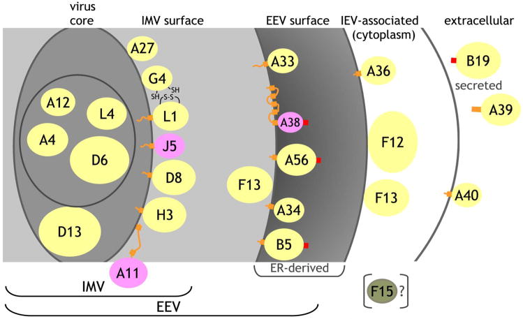
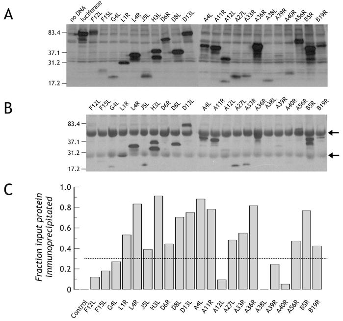
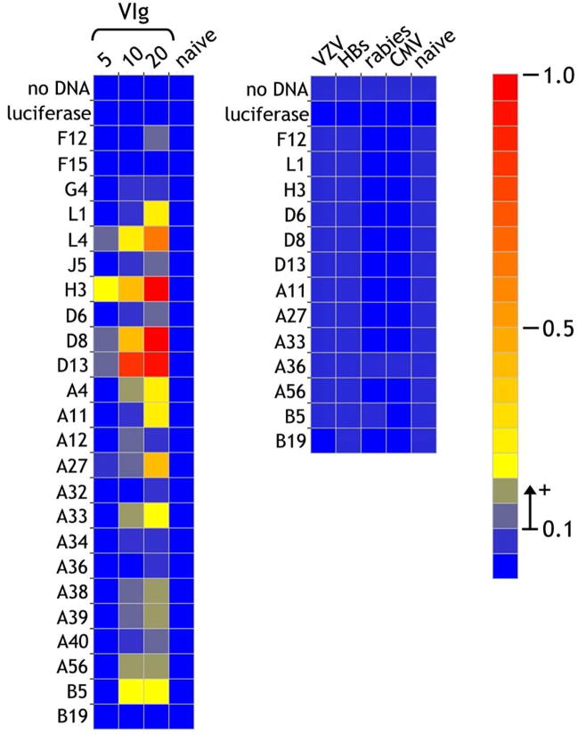
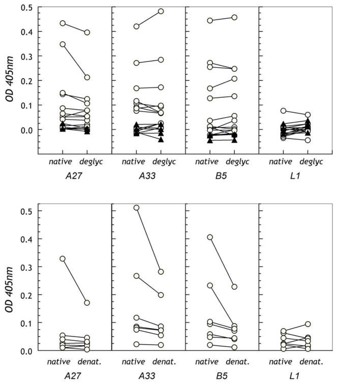
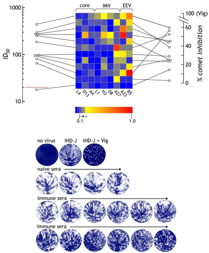
The renewed interest in strategies to combat infectious agents with epidemic potential has led to a re-examination of vaccination protocols against smallpox. To help define which antigens elicit a human antibody response, we have targeted proteins known or predicted to be presented on the surface of the intracellular mature virion (IMV) or the extracellular enveloped virion (EEV). The predicted ectodomains were expressed in a mammalian in vitro coupled transcription/translation reaction using tRNA(lys) precharged with lysine-epsilon-biotin followed by solid phase immobilization on 384-well neutravidin-coated plates. The generated array is highly specific and sensitive in a micro-ELISA format. By comparison of binding of vaccinia-immune sera to the reticulocyte lysate-produced proteins and to secreted post-translationally modified proteins, we demonstrate that for several proteins including the EEV proteins B5 and A33, proper recognition is dependent upon appropriate folding, with little dependence upon glycosylation per se. We further demonstrate that the humoral immune response to vaccinia among different individuals is not uniform in specificity or strength, as different IMV and EEV targets predominate within the group of immunogenic proteins. This heterogeneity likely results from the diversity of HLA Class II alleles and CD4 T helper cell epitopes stimulating B cell antibody production. Our findings have important implications both for design of new recombinant subunit vaccines as well as for methods of assaying the human antibody response utilizing recombinant proteins produced in vitro.


Figures

Figure 1
Figure 1. Virus topology of vaccinia proteins used for array
Based on prior publications and bioinformatics analysis (Table S1), proteins were assigned to the virus core, to the inner membrane of the IMV, to the IMV surface, to the EEV surface, to the outer membrane of the IEV, or as secreted from the host cell. For each protein, the yellow (experimentally demonstrated topology), pink (predicted) or green (F15) ellipse represents the domain synthesized and used in the array where the colored areas are proportional to the spherical size of each protein relative to the others. Not included in the constructed proteins are cytoplasmic domains or short inter-transmembrane regions (orange lines), transmembrane domains (orange blocks), or signal peptides (red blocks). A11 partitions predominantly in the cell but some may associate with the IMV, where the two indicated predicted transmembrane regions do not insert in the IMV membrane. The topology of A38 places it as a Type I integral membrane protein facing into the ER but it has not been unequivocally demonstrated to be on the EEV surface. A39 is secreted in the Copenhagen strain, but is membrane-bound in the vaccinia WR strain. B19 is secreted from the host cell. F15 is not part of the IMV but its topology is uncertain.
Figure 2
Figure 2. Production of vaccinia proteins and their immunoprecipitation with VIg
A. Production of biotin-labeled vaccinia protein domains. The desired vaccinia domains were produced incorporating tRNAlys precharged with ε-amino-biotinylated lysine. Synthesized proteins were detected with streptavidin-horse radish peroxidase. B. Immunoprecipitation of synthetically labeled proteins by human anti-vaccinia antibody. Identical amounts of protein to those loaded and detected in panel A were incubated with human anti-vaccinia hyperimmune IgG (VIg) and bound biotinylated protein was immunoprecipitated using Protein G-agarose. Following gel electrophoresis and transfer, bound protein was detected as in panel A. Arrows represent human Ig H and L chains, respectively. C. Broad reactivity of VIg to the synthetic antigen array. For each protein in panel A and panel B, the band relative intensity was normalized to luciferase run on the same gel. Since identical amounts of luciferase were run on each gel, the ratio of immunoprecipitated protein (panel B) to total protein loaded (panel A) gives a broad measure of VIg reactivity to specific antigens. Using a conservative cut-off threshold of 0.35 (indicated by the dotted line, strong reactivity is seen to L1, L4, J5, H3, D6, D8, D13, A4, A11, A27, A33, A36, A56, B5 and B19. Control is luciferase precipitated by VIg.
Figure 3
Figure 3. Purification of His(6)-tagged proteins and reactivity on nitrocellulose
A. Purification of vaccinia proteins synthesized in vitro. Reticulocyte lysate containing the newly-synthesized proteins were incubated with MagZ magnetic beads to bind the His(6)-tagged proteins. After washing and elution, the depleted lysate, eluate, or beads were run on a reducing SDS-PAGE gel and biotinylated protein detected by binding of streptavidin-peroxidase. B. Immobilization on nitrocellulose may affect the presentation of antigenic epitopes. Recombinant biotin-labeled H3, L1 and B5 vaccinia proteins purified by His-tag affinity were spotted in quadruplicate on nitrocellulose and probed with anti-myc-HRP directed against the C-terminal myc tag followed by chemiluminescent assay (right-hand panel). The same proteins were blotted in duplicate (10ng/spot) on a separate blot, incubated with VIg and detected with anti-human IgG-HRP.
Figure 4
Figure 4. Detection of VIg antigenic reactivities using the ELISA protein array
Vaccinia proteins produced by in transcription/translation in vitro were immobilized on neutravidin-coated plates through the biosynthetically-incorporated biotin tags. In the left-hand panel, VIg was incubated at 5, 10 and 20μg/ml and bound antibody detected by binding of anti-human IgG alkaline phospahatase activity. The highest concentration of 20μg/ml corresponds to a dilution of 1/2,500 from the stock. Immunoglobulin (20μg/ml) from a naïve individual was included as a control. As a further control for specificity, IgG from sera of individuals hyperimmunized against other microorganisms [varicella zoster (VZV), Hepatitis B secreted antigen (HBs), rabies and cytomegalovirus (CMV)] were also tested at 20μg/ml. The scale for the heatmap indicates optical density at 405nm detected in the ELISA assay. A value of >0.1 O.D. units above background was considered as significantly positive, while responses of 0.05–0.1 were considered borderline.
Figure 5
Figure 5. Sera background binding is an intrinsic property of IgG
A. Specific response, following background subtraction, to a subset of recombinant vaccinia antigens on the micro-ELISA array using sera from a vaccinated individual (1/300). B. Following Cibacron Blue (F3GA)-agarose treatment of the sera to remove albumin, specific responses with background subtracted are also reduced indicating partial removal of the IgG component. C. Following pretreatment of the sera with Protein A-agarose removing the IgG component, all specific anti-vaccinia protein reactivity is removed. D. For this individual, the background independent of any antigen presence was high; removal of albumin reduced the background by almost 0.5, but reduced the IgG signal by a similar amount (see panel B). Removal of the IgG component alone eliminated the background entirely.
Figure 6
Figure 6. Detection of anti-vaccinia reactivity from naïve, primary immunized, and immunized and boosted individuals
A. Purified IgG at 20μg/ml from 4 naïve individuals, 9 immunized individuals and 11 individuals vaccinated in childhood and subsequently boosted were assessed for vaccinia protein reactivity on the micro-ELISA array. Of the boosted individuals, one was tested more than 7 years after the boost and two were tested approximately 1 year after boost. All other vaccinated and boosted individuals were tested one to two months after the last exposure. VIg at 10μg/ml was used as a standard positive control. The lowest 4 rows separated from the main array indicate results for the same IgG preparations against recombinant A27, A33, B5 and L1 produced as secreted post-translationally-modified proteins by insect cells. All 4 antigens were immobilized at 200ng/well on uncoated plates followed by blocking with TBS-BSA (1%), after which the procedure was identical to that used for the reticulocyte lysate-produced proteins. B. The individuals tested at 1–2 months following boost were retested using IgG at 100μg/ml.
Figure 7
Figure 7. Optimal antigenic reactivity of sera requires post-translationally-modified or properly-folded target proteins
A. Untreated or endoglycosidase F1-treated proteins were immobilized on uncoated 384-well micro-ELISA plates and reactivity of purified IgG was assessed. B. Untreated or TFA-treated proteins were immobilized on ELISA plates for testing of reactivity of IgG from selected vaccinated individuals. In both panels, the dotted line indicates the cut-off for positivity where 0.05–0.1 is considered borderline and above 0.1 significantly positive.
Figure 8
Figure 8. The human antibody response to vaccinia target antigens is individually diverse but still leads to effective neutralization titers
Based on data in Figure 6, the responses of vaccinees to the dominantly recognized antigens are depicted, with each representing one individual. For each individual, the responses are connected by a line to the left indicating the corresponding ID50 for vaccinia infection (VV:Luc) of HeLa cells representing IMV neutralization. The red dotted line (ID50 = 20) represents background, where 20–30 ID50 is considered borderline and >30 significantly positive. To the right of each row, a line connects to the % inhibition of comet formation representing EEV activity. Using image densitometry, the VIg responses were set to 100% inhibition and the responses to naïve sera were used to set the zero baseline. The presumed virus topology of each antigen is indicated at the top of array. In the lower panel, the representative comet inhibition responses are depicted where the 12 immune sera are ordered in increasing comet inhibition activity following incubation of BSC-40 cells with IHD-J EEV-forming virus (100pfu).

References

    1. Fauci AS. Emerging infectious diseases: a clear and present danger to humanity. JAMA. 2004 Oct 20;292(15):1887–8. - PubMed
    1. Morens DM, Folkers GK, Fauci AS. The challenge of emerging and re-emerging infectious diseases. Nature. 2004 Jul 8;430(6996):242–9. - PMC - PubMed
    1. Fauci AS, Touchette NA, Folkers GK. Emerging infectious diseases: a 10-year perspective from the National Institute of Allergy and Infectious Diseases. Emerg Infect Dis. 2005 Apr;11(4):519–25. - PMC - PubMed
    1. Lane JM, Goldstein J. Evaluation of 21st-century risks of smallpox vaccination and policy options. Ann Intern Med. 2003 Mar 18;138(6):488–93. - PubMed
    1. Rotz LD, Dotson DA, Damon IK, Becher JA. Vaccinia (smallpox) vaccine: recommendations of the Advisory Committee on Immunization Practices (ACIP), 2001. MMWR Recomm Rep. 2001 Jun 22;50(RR10):1–25. quiz CE1–7. - PubMed

Publication types