Review
. 2008:85:225-60.
doi: 10.1016/S0070-2153(08)00809-0.

The primary cilium at the crossroads of mammalian hedgehog signaling

Affiliations
Review

The primary cilium at the crossroads of mammalian hedgehog signaling

Sunny Y Wong et al. Curr Top Dev Biol. 2008.

Abstract

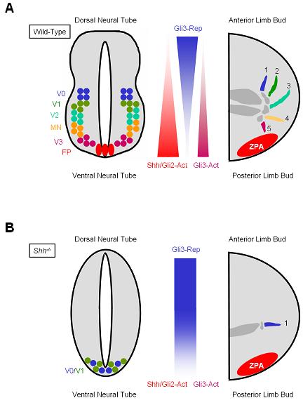
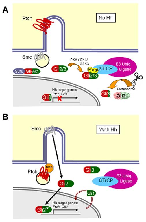
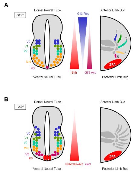
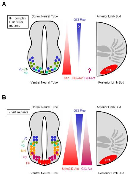
Cilia function as critical sensors of extracellular information, and ciliary dysfunction underlies diverse human disorders including situs inversus, polycystic kidney disease, retinal degeneration, and Bardet-Biedl syndrome. Importantly, mammalian primary cilia have recently been shown to mediate transduction of Hedgehog (Hh) signals, which are involved in a variety of developmental processes. Mutations in several ciliary components disrupt the patterning of the neural tube and limb bud, tissues that rely on precisely coordinated gradients of Hh signal transduction. Numerous components of the Hh pathway, including Patched, Smoothened, and the Gli transcription factors, are present within primary cilia, indicating that key steps of Hh signaling may occur within the cilium. Because dysregulated Hh signaling promotes the development of a variety of human tumors, cilia may also have roles in cancer. Together, these findings have shed light on one mechanism by which primary cilia transduce signals critical for both development and disease.


Figures

Figure 1
Figure 1. Proper patterning of the neural tube and limb buds is mediated by a gradient of Shh
(A) In the neural tubes of wild-type embryos, Shh produced by the notochord, floor plate (FP) and possibly gut forms a gradient along the dorsal-ventral axis. Similarly, Shh released by cells in the zone of polarizing activity (ZPA) in the limb buds forms a gradient along the anterior-posterior axis. Increased Shh concentration and duration of action are associated with increased Gli2 activator (Gli2-Act) and Gli3 activator (Gli3-Act) functions, and reduced Gli3 repressor (Gli3-Rep) formation. High level Hh signaling is critical for specifying the ventral-most cell types in the neural tube, including FP and V3 interneurons (V3). More dorsal cell types (V0, V1, V2 and motorneurons (MN)) are specified by lower levels of Hh signaling. In limb buds, the Shh and Gli3-Rep gradients are interpreted for the specification of digit identity (digits 1-5). (B) In Shh mutant embryos, the gradient of Hh pathway activation is absent. Gli3 is mostly converted into Gli3-Rep along the dorsal-ventral axis of the neural tube and the anterior-posterior axis of the limb buds, although Ihh may still activate the pathway to some degree. In mutant neural tubes, only the V0 and V1 neuronal subtypes are specified, and in mutant limb buds, only the anterior-most digit (digit 1) is formed.
Figure 2
Figure 2. A model of mammalian Hh signal transduction
(A) In the absence of Shh, Ptch localizes to the primary cilium and, through an unknown mechanism, prevents Smo from entering the cilium. Gli2 and Gli3 are phosphorylated by kinases, including PKA, CKI and GSK3, to generate phosphopeptide binding motifs for β-TrCP, which recruits an E3 ubiquitin ligase complex. Ubiquitination of Gli2 and Gli3 targets these proteins to the proteasome, which processes Gli3 into a carboxy-terminal-truncated repressor form and degrades Gli2. Gli3 subsequently enters the nucleus and inhibits transcription of downstream Hh target genes such as Ptch1 and Gli1. (B) In the presence of Shh, Ptch is internalized, allowing Smo to traffic into the primary cilium. It is unclear whether Smo traffics directly to the cilium, or accumulates first in the plasma membrane. At the cilium, Smo inhibits the formation of Gli3 repressor and activates Gli2, which enters the nucleus to promote transcription of Ptch1 and Gli1, whose protein products negatively and positively feed back on this pathway, respectively.
Figure 3
Figure 3. The neural tube and limb buds differ in their reliance on Gli2 and Gli3 for proper patterning
(A) In Gli2 mutant embryos, gradients of Shh and Gli3-Rep/Act are established, but downstream signaling is not properly activated. In the neural tube, which relies predominantly on Gli2 activator function, specification of neuronal subtypes that require the highest levels of Hh pathway activity (FP and V3) is deficient. Because proper patterning of the limb bud depends upon a gradient of Gli3-Act/-Rep, and not on Gli2-Act, digit specification is normal in Gli2 mutants. (B) In Gli3 mutant embryos, gradients of Shh and Gli2-Act are established, but formation of Gli3-Rep is absent. Gli3-deficient neural tubes display only a subtle expansion of V0 and V1 neuronal subtypes. Because proper limb patterning depends on a gradient of Gli3-Rep, Gli3 mutant limb buds upregulate genes normally repressed by Gli3-Rep, including Gremlin and Hoxd13, and exhibit polydactyly.
Figure 4
Figure 4. Many IFT mutations cause polydactyly and loss of ventral neural tube fates
(A) In embryos lacking IFT components such as IFT88, IFT172 and Kif3a, a gradient of Shh is established, but signaling through Gli2-Act and formation of Gli3-Rep are disrupted. Consequently, these mutants do not properly activate the Hh transcriptional targets, and display loss of ventral cell types (FP, V3, MN) and expansion of more dorsal subtypes (V0, V1, V2) in the neural tube. In IFT mutant limb buds, disruption of Gli3-Rep formation leads to polydactyly. (B) In contrast to other IFT mutants, THM1 mutants display increased Hh signaling mediated by Gli2-Act and Gli3-Act, leading to an increase in ventral subtypes (FP, V3, MN) in the neural tube. THM1 is also essential for Gli3 processing, defects of which in THM1 mutants result in polydactyly.
Figure 5
Figure 5. The primary cilium likely coordinates a variety of signaling pathways important for cancer
In Hh-dependent cancers such as BCC and medulloblastoma, the cilium may modulate the activity of signaling pathways in addition to Hh. For instance, localization of PDGF receptor-αα to the cilium is important for transducing downstream signals mediated by Akt and MAP kinases. The cilium may also normally restrain Wnt pathway activity by preventing the accumulation of β-catenin. The balance of the downstream effects of these pathways likely governs cellular decisions for proliferation and apoptosis.

References

    1. Adams NA, Awadein A, Toma HS. The retinal ciliopathies. Ophthalmic Genet. 2007;28:113–125. - PubMed
    1. Ahn S, Joyner AL. Dynamic changes in the response of cells to positive hedgehog signaling during mouse limb patterning. Cell. 2004;118:505–516. - PubMed
    1. Ansley SJ, Badano JL, Blacque OE, Hill J, Hoskins BE, Leitch CC, Kim JC, Ross AJ, Eichers ER, Teslovich TM, Mah AK, Johnsen RC, Cavender JC, Lewis RA, Leroux MR, Beales PL, Katsanis N. Basal body dysfunction is a likely cause of pleiotropic Bardet-Biedl syndrome. Nature. 2003;425:628–633. - PubMed
    1. Arima T, Shibata Y, Yamamoto T. A deep-etching study of the guinea pig tracheal cilium with special reference to the ciliary transitional region. J Ultrastruct Res. 1984;89:34–41. - PubMed
    1. Aszterbaum M, Epstein J, Oro A, Douglas V, LeBoit PE, Scott MP, Epstein EH., Jr Ultraviolet and ionizing radiation enhance the growth of BCCs and trichoblastomas in patched heterozygous knockout mice. Nat Med. 1999;5:1285–1291. - PubMed

Publication types

Substances

LinkOut - more resources