Review
. 2008:85:303-32.
doi: 10.1016/S0070-2153(08)00811-9.

Cilia involvement in patterning and maintenance of the skeleton

Affiliations
Review

Cilia involvement in patterning and maintenance of the skeleton

Courtney J Haycraft et al. Curr Top Dev Biol. 2008.

Abstract

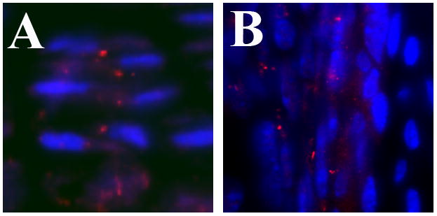
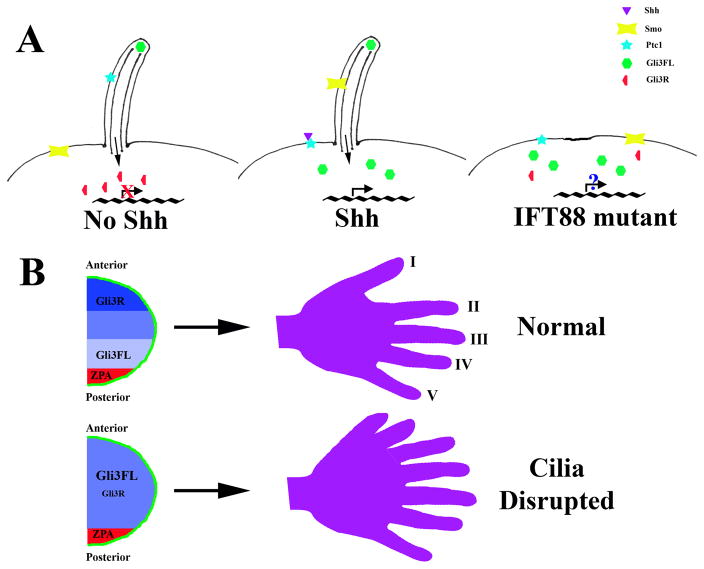
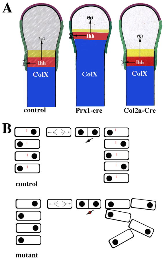
Although the expression of cilia on chondrocytes was described over 40 years ago, the importance of this organelle in skeletal development and maintenance has only recently been recognized. Primary cilia are found on most mammalian cells and have been shown to play a role in chemosensation and mechanosensation. A growing number of human pleiotropic syndromes have been shown to be associated with ciliary or basal body dysfunction. Skeletal phenotypes, including alterations in limb patterning, endochondral bone formation, craniofacial development, and dentition, have been described in several of these syndromes. Additional insights into the potential roles and mechanisms of cilia action in the mammalian skeleton have been provided by research in model organisms including mouse and zebrafish. In this article we describe what is currently known about the localization of cilia in the skeleton as well as the roles and underlying molecular mechanisms of cilia in skeletal development.


Figures

Figure 1
Figure 1. Localization of cilia on chondrocytes and perichondrial cells
(A) Cilia, visualized by immunostaining with anti-acetylated α-tubulin antibodies (red), are aligned in the center of columns of proliferating chondrocytes in the growth plate of endochondral bones. Nuclei (blue) are located on alternate sides of the cells within a column. (B) In the perichondrium, cilia are present on both elongated and cuboidal cells adjacent to the cartilage anlagen (right side of image). Cilia were visualized by immunostaining with anti-Ift88 antibodies (red). Nuclei are blue.
Figure 2
Figure 2. Mammalian Hh signal transduction and limb patterning
(A) Hh signal transduction. In the absence of Hh ligand, Ptc1 localizes to the ciliary membrane. Full length Gli3 (Gli3FL) is found at the distal tip of the cilium and subsequent proteolytic processing results in increased amounts of Gli3 repressor (Gli3R). Gli3R is a potent transcriptional repressor resulting in repression of target gene transcription including Ptc1 and Gli1. Upon ligand binding, Ptc1 translocates out of the cilium and Smo is localized in the cilium membrane. Smo translocation to the cilium inhibits Gli3 processing leading to an increased Gli3FL:Gli3R ratio allowing transcription of target genes. In Ift88 mutants, the cilium is not formed and Gli3 processing is inefficient. As a result, target genes such as Gremlin and Hand2, which are repressed by Gli3R, are expressed while transcription of Gli1 and Ptc1 is not activated likely due to disruption of Gli2 function by an unknown mechanism. (B) Role of Shh in development of the mammalian limb. In the developing limb, Shh is expressed by cells in the ZPA (red). Activation of Shh signaling leads to decreased Gli3R levels in the posterior limb bud while the level of Gli3R remains high in the anterior limb bud. In cilia mutants, such as those lacking Ift88 or Kif3a, disruption of the normal gradient of Gli3FL:Gli3R in conjunction with loss of Gli2 activity across the limb bud results in polydactyly.
Figure 3
Figure 3. A role for primary cilia in endochondral bone formation and the post natal growth plate
(A) Primary cilia regulate Ihh signaling during embryonic endochondral bone formation. Ihh is expressed in chondrocytes that are committed to hypertrophic differentiation (red area). Ihh normally acts on both prehypertrophic chondrocytes (gray and yellow areas) and the perichondrium (green). Ptc1, a downstream target of Ihh signaling, was used to determine how depletion of cilia affects Ihh signaling during endochondral bone formation. In normal control mice, cilia are present on chondrocytes as well as cells in the perichondrium and Ptc1 expression is detected in both cell types. In mice in which ciliogenesis is disrupted in Prx1-Cre expressing cells, cilia are absent from both chondrocytes and cells in the perichondrium. Disruption of ciliogenesis in the cartilage and in the perichondrium resulted in accelerated hypertrophic differentiation (blue area) similar to that seen in the Ihh-null mice. Ptc1 expression was dramatically reduced in both chondrocytes and cells of the perichondrium confirming that Ihh signaling was disrupted. In contrast, when ciliogenesis was disrupted using Col2a-Cre, which only targets the chondrocytes, hypertrophic differentiation was normal. Ptc1 expression was reduced in chondrocytes but maintained in the perichondrium. (B) Primary cilia mediate chondrocyte rotation in the post-natal growth plate. In normal control mice, flat cells in the growth plate have polarity and are aligned in columns parallel to the long-axis of the bone. Cell division occurs perpendicular to the long axis. One cell then migrates under the other to form the columns. This process is called chondrocyte rotation. Cilia are present on chondrocytes in the columns and are required for normal chondrocyte rotation. In cilia depleted growth plates, chondrocytes still divide perpendicular to the long axis of the bone and polarity is maintained; however, the orientation of the cells one to another is altered and the cells are not maintained in columns.

References

    1. Alvarez J, Sohn P, Zeng X, Doetschman T, Robbins DJ, Serra R. Development. 2002;129:1913–24. - PubMed
    1. Ascenzi MG, Lenox M, Farnum C. J Struct Biol. 2007;158:293–306. - PMC - PubMed
    1. Aszodi A, Hunziker EB, Brakebusch C, Fassler R. Genes Dev. 2003;17:2465–79. - PMC - PubMed
    1. Beales PL, Bland E, Tobin JL, Bacchelli C, Tuysuz B, Hill J, Rix S, Pearson CG, Kai M, Hartley J, Johnson C, Irving M, Elcioglu N, Winey M, Tada M, Scambler PJ. Nat Genet. 2007;39:727–9. - PubMed
    1. Bisgrove BW, Yost HJ. Development. 2006;133:4131–43. - PubMed

LinkOut - more resources