Comparative Study
. 2009 Mar;11(3):246-55.
doi: 10.1093/eurjhf/hfn027. Epub 2009 Jan 12.

Enhanced myogenic constriction of mesenteric artery in heart failure relates to decreased smooth muscle cell caveolae numbers and altered AT1- and epidermal growth factor-receptor function

Affiliations
Comparative Study

Enhanced myogenic constriction of mesenteric artery in heart failure relates to decreased smooth muscle cell caveolae numbers and altered AT1- and epidermal growth factor-receptor function

Ying Xu et al. Eur J Heart Fail. 2009 Mar.

Abstract

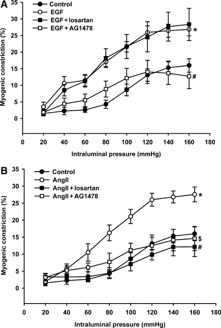
Aims: We previously showed that enhanced myogenic constriction (MC) of peripheral resistance arteries involves active AT(1) receptors in chronic heart failure (CHF). Recent data suggest both transactivation of EGF receptors and caveolae-like microdomains to be implicated in the activity of AT(1) receptors. Thus, we assessed their roles in increased MC in mesenteric arteries of CHF rats.

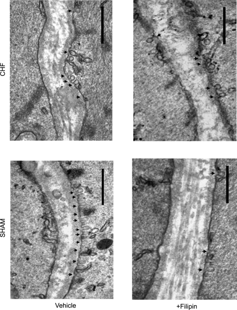
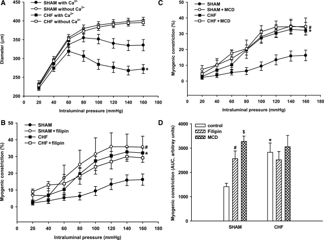
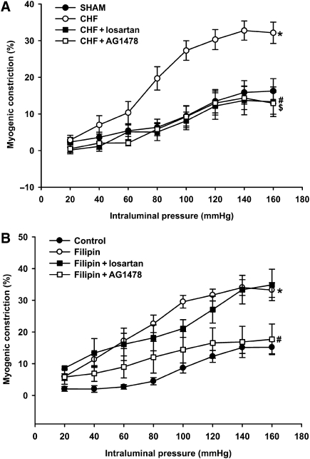
Methods and results: Male Wistar rats underwent myocardial infarction to induce CHF and were sacrificed after 12 weeks. The number of caveolae in smooth muscle cells (SMC) of mesenteric arteries of CHF rats was decreased by 43.6 +/- 4.0%, this was accompanied by increased MC, which was fully normalized to the level of sham by antagonists of the AT(1)-receptor (losartan) or EGF-receptor (AG1478). Acute disruption of caveolae in sham rats affected caveolae numbers and MC to a similar extent as CHF, however MC was only reversed by the antagonist of the EGF-receptor, but not by the AT(1)-receptor antagonist. Further, in sham rats, MC was increased by a sub-threshold concentration of angiotensin II and reversed by both AT(1)- as well as EGF-receptor inhibition. In contrast, increased MC by a sub-threshold concentration of EGF was only reversed by EGF receptor inhibition.

Conclusion: These findings provide the first evidence that decreased SMC caveolae numbers are involved in enhanced MC in small mesenteric arteries, by affecting AT(1)- and EGF-receptor function. This suggests a novel mechanism involved in increased peripheral resistance in CHF.


Figures

Figure 1
Figure 1
Visualization of caveolae in mesenteric artery vascular smooth muscle cells (VSMC) of sham rats and myocardial infarction rats with chronic heart failure (CHF) using transmission electron microscopy. Representative sections treated either with vehicle or with filipin are shown. Arrowheads indicate caveolae; bar, 0.5 µm in each case.
Figure 2
Figure 2
Immunohistochemistry for caveolin-1 and caveolin-3. Representative pictures of sections from whole rings of mesenteric artery stained for caveolin-1 (A) and caveolin-3 (B) (bars represent 25 µm, arrows indicate positive smooth muscle cells). Left panel shows negative staining in control Ig treated sections. (C) Quantification of the caveolin-1 and caveolin-3 staining selectively in the vascular smooth muscle layer. Data are expressed as mean±SEM (n = 6 in sham and n = 4 in CHF).
Figure 3
Figure 3
Myogenic constriction determined from pressure–diameter relationships in small mesenteric arteries of sham rats and myocardial infarction rats with chronic heart failure (n = 15 for both groups). (A) Arteries were subjected to stepwise increase in intraluminal pressure in the absence and presence of extracellular calcium (Ca2+) and the diameter was determined at each step. Active diameters in the presence of Ca2+—but not passive diameters in the absence of Ca2+—were significantly smaller in the arteries of rats with CHF (*P < 0.05 vs. sham with Ca2+ in A). (B and C) Consequently, myogenic constriction (% of passive diameter) was significantly increased in arteries of rats with CHF (*P < 0.05 vs. sham in B and C). Treatment with filipin (4 µg mL−1, n = 6) or methyl-β-cyclodextrin (MCD, 1 mmol L−1, n = 6) significantly increased myogenic constriction in arteries of sham rats (#P < 0.05 vs. sham in B and C, respectively), i.e. to levels similar as those observed in arteries of rats with CHF. (D) Similar results were obtained with endothelium-denuded arteries, indicating that the augmenting effect of filipin and MCD on myogenic constriction (represented by the AUC bar-size) of sham arteries was endothelium-independent (*,#,$P < 0.05 for CHF, sham filipin, and sham MCD, respectively, vs. sham control). Data are mean±SEM (n = 6–12 in each condition).
Figure 4
Figure 4
Effect of EGF- and AT1-receptor blockade on increased myogenic constriction in mesenteric artery resulting from CHF or caveolae disruption. (A) Increased myogenic constriction in CHF (*P < 0.05 vs. sham) was fully normalized in the presence of the AT1-receptor antagonist losartan (10 µmol L−1,#P < 0.05 vs. CHF) or the EGF-receptor inhibitor AG1478 (5 µmol L−1, $P < 0.05 vs. CHF). (B) Treatment with filipin significantly increased myogenic constriction in arteries of sham rats (*P < 0.05); the EGF-receptor inhibitor, AG1478, significantly reduced myogenic constriction in filipin-treated sham arteries of sham rats (#P < 0.05 vs. filipin). In contrast, losartan did not affect enhanced myogenic constriction after filipin. Data are mean±SEM (n = 6–14 in each condition).
Figure 5
Figure 5
Augmentation of the myogenic response in normal mesenteric artery preparations of sham rats by sub-threshold concentrations of EGF (50 ng mL−1; *P < 0.05 vs. sham control; A) and angiotensin II (AngII, 10 nmol L−1; *P < 0.05 vs. sham control; B). (A) Enhanced myogenic constriction in the presence of EGF was selectively normalized by additional presence of AG1478 (5 µmol L−1, #P < 0.05 vs. EGF), but not by losartan (10 µmol L−1). (B) Enhanced myogenic constriction in the presence of angiotensin II was normalized both by losartan as well as AG1478 (#,$P < 0.05 for AngII + losartan and AngII + AG1478, respectively, vs. AngII only). Data are mean ± SEM (n = 6–13 in each condition).
Figure 6
Figure 6
Co-localization of the EGF- and AT1-receptors. Immunofluorescent staining was performed for EGF receptor (A, green, arrows) and AT1 receptor (B, red, arrows) on mesenteric arteries and analysed with confocal microscopy. Co-localization is shown in yellow (C, arrows). Bar indicates 20 µm.

Similar articles

Cited by

References

    1. Schrier RW, Abraham WT. Mechanisms of disease - Hormones and hemodynamics in heart failure. N Eng J Med. 1999;341:577–585. - PubMed
    1. Davis MJ, Hill MA. Signaling mechanisms underlying the vascular myogenic response. Physiol Rev. 1999;79:387–423. - PubMed
    1. Gschwend S, Henning RH, Pinto YM, de Zeeuw D, van Gilst WH, Buikema H. Myogenic constriction is increased in mesenteric resistance arteries from rats with chronic heart failure: instantaneous counteraction by acute AT(1) receptor blockade. Br J Pharmacol. 2003;139:1317–1325. - PMC - PubMed
    1. Kagiyama S, Eguchi S, Frank GD, Inagami T, Zhang YC, Phillips MI. Angiotensin II-induced cardiac hypertrophy and hypertension are attenuated by epidermal growth factor receptor antisense. Circulation. 2002;106:909–912. - PubMed
    1. Ushio-Fukai M, Hilenski L, Santanam N, Becker PL, Ma Y, Griendling KK, Alexander RW. Cholesterol depletion inhibits epidermal growth factor receptor transactivation by angiotensin II in vascular smooth muscle cells: role of cholesterol-rich microdomains and focal adhesions in angiotensin II signaling. J Biol Chem. 2001;276:48269–48275. - PubMed

Publication types

MeSH terms

Substances