. 2009 Mar 20;284(12):7820-31.
doi: 10.1074/jbc.M809332200. Epub 2009 Jan 15.

The C-terminal region of laminin beta chains modulates the integrin binding affinities of laminins

Affiliations

The C-terminal region of laminin beta chains modulates the integrin binding affinities of laminins

Yukimasa Taniguchi et al. J Biol Chem. .

Abstract

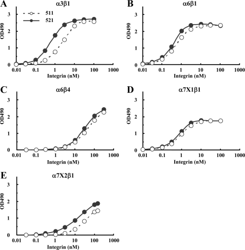
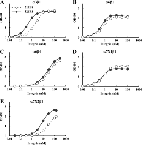
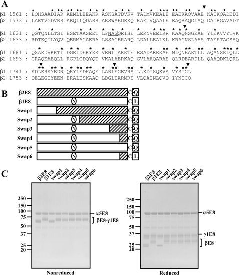
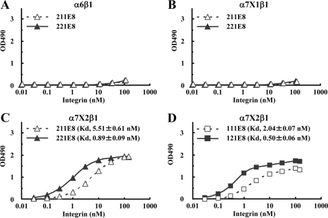
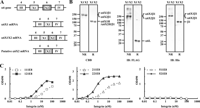
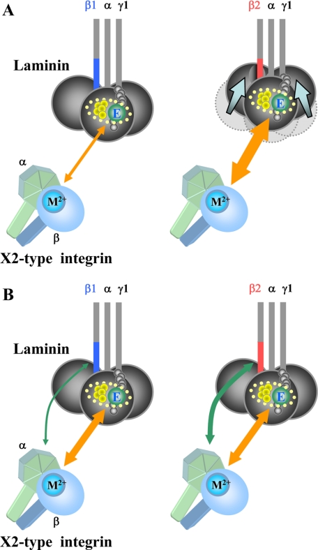
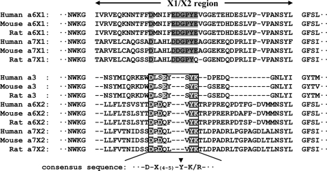
Laminins are major cell-adhesive proteins in basement membranes that are capable of binding to integrins. Laminins consist of three chains (alpha, beta, and gamma), in which three laminin globular modules in the alpha chain and the Glu residue in the C-terminal tail of the gamma chain have been shown to be prerequisites for binding to integrins. However, it remains unknown whether any part of the beta chain is involved in laminin-integrin interactions. We compared the binding affinities of pairs of laminin isoforms containing the beta1 or beta2 chain toward a panel of laminin-binding integrins, and we found that beta2 chain-containing laminins (beta2-laminins) bound more avidly to alpha3beta1 and alpha7X2beta1 integrins than beta1 chain-containing laminins (beta1-laminins), whereas alpha6beta1, alpha6beta4, and alpha7X1beta1 integrins did not show any preference toward beta2-laminins. Because alpha3beta1 contains the "X2-type" variable region in the alpha3 subunit and alpha6beta1 and alpha6beta4 contain the "X1-type" region in the alpha6 subunit, we hypothesized that only integrins containing the X2-type region were capable of discriminating between beta1-laminins and beta2-laminins. In support of this possibility, a putative X2-type variant of alpha6beta1 was produced and found to bind preferentially to beta2-laminins. Production of a series of swap mutants between the beta1 and beta2 chains revealed that the C-terminal 20 amino acids in the coiled-coil domain were responsible for the enhanced integrin binding by beta2-laminins. Taken together, the results provide evidence that the C-terminal region of beta chains is involved in laminin recognition by integrins and modulates the binding affinities of laminins toward X2-type integrins.


Figures

FIGURE 1.
FIGURE 1.
SDS-PAGE and immunoblot analyses of purified laminin-511 and -521. A and B, purified laminin-511 and -521 (labeled β1 and β2, respectively) were analyzed by SDS-PAGE in 4% gels under nonreducing or reducing conditions. The separated proteins were visualized by Coomassie Brilliant Blue staining (A) or transferred onto polyvinylidene difluoride membranes followed by immunoblotting (IB) with mAbs against theα5 (15H5), β1 (DG10), β2 (6A3) and, γ1 (C12SW) chains (B). The positions of molecular size markers, including nonreduced and reduced Engelbreth-Holm-Swarm-laminin (800 and 400 kDa, respectively), are shown in the left margin. The asterisk denotes the laminin α5 chain lacking LG4–5.
FIGURE 2.
FIGURE 2.
Titration curves of recombinant integrins bound to laminin-511 and -521. A–E, increasing concentrations of recombinant α3β1(A), α6β1(B), α6β4(C), α7X1β1(D), and α7X2β1(E) integrins were allowed to bind to microtiter plates coated with laminin-511 (open circles) or laminin-521 (closed circles) in the presence of 1 mm MnCl2. Bound integrins were quantified using an antibody against the ACID/BASE peptides attached to the C termini of the α and β integrin subunits. The amounts of the integrins bound in the presence of 10 mm EDTA were taken as nonspecific binding and subtracted as background. The results shown are the means of duplicate determinations. The apparent dissociation constants of the recombinant integrins are summarized in Table 1.
FIGURE 3.
FIGURE 3.
SDS-PAGE and immunoblot analyses of purified laminin-511E8 and -521E8. A, schematic diagrams of a full-length laminin and a recombinant E8 fragment. The cysteine (C) residues conserved in the C termini of the laminin β and γ chains are indicated. B and C, laminin-511E8 and -521E8 (labeled β1 and β2, respectively) were subjected to SDS-PAGE in 5–20% gradient polyacrylamide gels, followed by Coomassie Brilliant Blue staining (B) or immunoblotting (IB) with specific antibodies against the α5E8 (anti-His5), β1E8/β2E8 (anti-HA), and γ1E8 (anti-FLAG) fragments (C) under nonreducing (NR) and reducing (R) conditions. The positions of molecular size markers are shown in the left margin.
FIGURE 4.
FIGURE 4.
Titration curves of recombinant integrins bound to laminin-511E8 and -521E8. A–E, increasing concentrations of recombinant α3β1(A), α6β1(B), α6β4(C), α7X1β1(D), and α7X2β1(E) integrins were allowed to bind to microtiter plates coated with laminin-511E8 (open circle) and -521E8 (closed circle) in the presence of 1 mm MnCl2. The amounts of the integrins bound in the presence of 10 mm EDTA were taken as nonspecific binding and subtracted as background. The results shown are the means of duplicate determinations. Apparent dissociation constants of recombinant integrins are summarized in Table 1.
FIGURE 5.
FIGURE 5.
Production of swap mutants between the laminin β1 and β2 chains. A, sequence alignment of the C-terminal regions of the human laminin β1-(1561–1786) and β2-(1573–1798) chains. The boxed amino acid sequence (Asn-Ala-Ser) is a putative N-linked glycosylation site. Conserved amino acids between the laminin β1 and β2 chains are indicated by asterisks. The boundaries between the β1 and β2 chains in the swap mutants are indicated by arrowheads. B, schematic representations of the β1/β2 swap mutants. The β1- and β2-derived sequences are represented by open boxes and hatched boxes, respectively. C, SDS-PAGE profiles of recombinant E8 fragments containing control and swapped βE8 chains were analyzed using 5–20% gradient polyacrylamide gels under nonreducing (left panel) and reducing (right panel) conditions, followed by Coomassie Brilliant Blue staining. The positions of molecular size markers are shown in the left margin.
FIGURE 6.
FIGURE 6.
Titration curves of recombinant integrins bound to laminin-111E8/121E8 and laminin-211E8/221E8. A–D, increasing concentrations of recombinant α6β1(A), α7X1β1(B), and α7X2β1(C and D) integrins were allowed to bind to microtiter plates coated with laminin-211E8 (open triangles) and -221E8 (closed triangles) (AC) and laminin-111E8 (open squares) and -121E8 (closed squares) (D) in the presence of 1 mm MnCl2. The amounts of the integrins bound in the presence of 10 mm EDTA were taken as nonspecific binding and subtracted as background. The results shown are the means of duplicate determinations. The apparent dissociation constants are expressed as the means ± S.D. of three independent experiments.
FIGURE 7.
FIGURE 7.
Ligand-binding specificities of recombinant α6X2β1 integrin. A, schematic diagrams of the exon/intron structures of the part of the human integrin α6 gene encompassing domains 4–7 and the RNA transcripts arising from alternative splicing at the X1/X2 exons. Exons 4–7 (open boxes or dotted boxes) encode the blade III, X1, X2, and blade IV regions, respectively. B, recombinant α6X1β1 and α6X2β1 integrins (labeled X1 and X2, respectively) were subjected to SDS-PAGE in 5–20% gradient gels under nonreducing (NR) and reducing (R) conditions, followed by Coomassie Brilliant Blue (CBB) staining (left panel) or immunoblotting with antibodies against the FLAG tag (for detection of the α6 light chain; middle panel) and His5 tag (for detection of the β1 subunit; right panel). The positions of molecular size markers are shown in the left margin. C, titration curves of recombinant α6X2β1 integrin bound to laminin-111E8/121E8, -211E8/221E8, and -511E8/521E8. Increasing concentrations of recombinant α6X2β1 integrins were allowed to bind to microtiter plates coated with laminin-111E8 (open squares), laminin-121E8 (closed squares), laminin-211E8 (open triangles), laminin-221E8 (closed triangles), laminin-511E8 (open circles), or laminin-521E8 (closed circles) in the presence of 1 mm MnCl2. The amounts of the integrins bound in the presence of 10 mm EDTA were taken as nonspecific binding and subtracted as background. The results shown are the means of duplicate determinations.
FIGURE 8.
FIGURE 8.
Models for the mechanism by which the laminin β2 chain potentiates the binding affinity toward X2-type laminin-binding integrins. A, interaction between the C-terminal region of the β2 chain and the LG1–3 modules of the α chain fine-tunes the conformation of LG1–3, which are assembled into a cloverleaf shape, and thereby up-regulates the binding affinities of β2-laminins for X2-type laminin-binding integrins. The items encircled with dotted lines are the putative integrin-binding site comprised of the glutamic acid (E) residue in the C-terminal region of the γ1 chain (green) and undefined residues within the LG1–3 modules of the α chain (yellow), the former coordinating the divalent metal ion (M2+) in the MIDAS motif of the integrin β subunit, whereas the latter possibly interacts with the β-propeller domain of the integrin α subunit. The differences in the affinities between the putative integrin-binding site and X2-type integrins are reflected by the width of the orange two-way arrows connecting them. B, C-terminal region of the laminin β2 chain interacts directly with X2-type integrins and thereby potentiates the binding affinities of β2-laminins for X2-type laminin-binding integrins. The C-terminal region of the β1 chain may also interact with X2-type integrins with significantly lower affinities than those of the β2 chain. The width of the green two-way arrows reflects the strength of the interaction between the C-terminal region of the β chains and X2-type integrins. The orange two-way arrows refer to the interaction between the integrin and the putative integrin-binding site of laminin comprised of the LG1–3 modules of the α chain and the C-terminal tail of the γ1 chain.
FIGURE 9.
FIGURE 9.
Multiple sequence alignment of the variable regions of the integrin α3, α6, and α7 subunits. The amino acid sequences of the X1 and X2 regions of the integrin α3, α6, and α7 subunits of different vertebrate species were aligned using ClustalX (35). The highly conserved sequence in the variable region of the X2-type integrins is boxed. The consensus residues in the conserved sequence are highlighted in black. The conserved sequence in the X1 region that corresponds to the consensus sequence in the X2 region is shaded. The arrowhead indicates the tyrosine residue equivalent to Tyr208 in the integrin α5 subunit.

Similar articles

Cited by

References

    1. Tunggal, P., Smyth, N., Paulsson, M., and Ott, M. C. (2000) Microsc. Res. Tech. 51 214-227 - PubMed
    1. Colognato, H., and Yurchenco, P. D. (2000) Dev. Dyn. 218 213-234 - PubMed
    1. Miner, J. H., and Yurchenco, P. D. (2004) Annu. Rev. Cell Dev. Biol. 20 255-284 - PubMed
    1. Smyth, N., Vatansever, H. S., Murray, P., Meyer, M., Frie, C., Paulsson, M., and Edgar, D. (1999) J. Cell Biol. 144 151-160 - PMC - PubMed
    1. Miner, J. H., Cunningham, J., and Sanes, J. R. (1998) J. Cell Biol. 143 1713-1723 - PMC - PubMed

Publication types

LinkOut - more resources