. 2009 Mar 27;284(13):8539-47.
doi: 10.1074/jbc.M808231200. Epub 2009 Jan 15.

Cytoglobin is expressed in the vasculature and regulates cell respiration and proliferation via nitric oxide dioxygenation

Affiliations

Cytoglobin is expressed in the vasculature and regulates cell respiration and proliferation via nitric oxide dioxygenation

Katharine E Halligan et al. J Biol Chem. .

Abstract

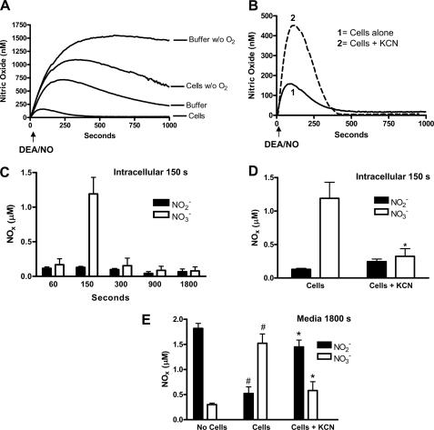
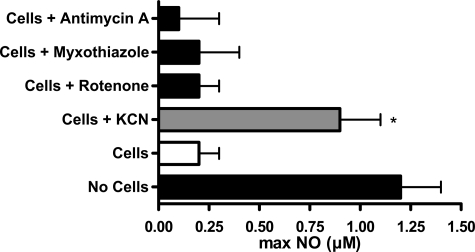
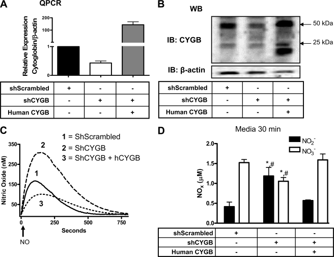
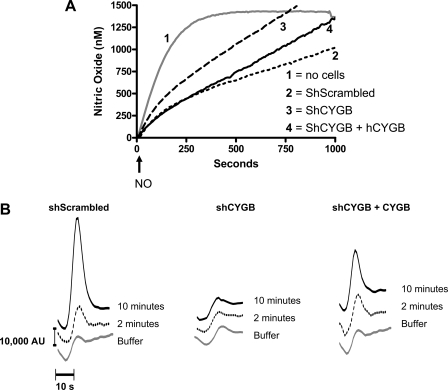
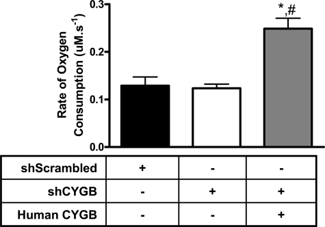
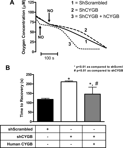
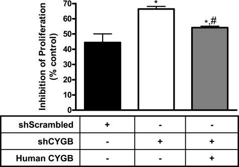
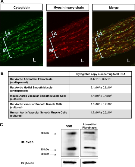
Disposition of the second messenger nitric oxide (NO) in mammalian tissues occurs through multiple pathways including dioxygenation by erythrocyte hemoglobin and red muscle myoglobin. Metabolism by a putative NO dioxygenase activity in non-striated tissues has also been postulated, but the exact nature of this activity is unknown. In the present study, we tested the hypothesis that cytoglobin, a newly discovered hexacoordinated globin, participates in cell-mediated NO consumption. Stable expression of small hairpin RNA targeting cytoglobin in fibroblasts resulted in decreased NO consumption and intracellular nitrate production. These cells were more sensitive to NO-induced inhibition of cell respiration and proliferation, which could be restored by re-expression of human cytoglobin. We also demonstrated cytoglobin expression in adventitial fibroblasts as well as vascular smooth muscle cells from various species including human and found that cytoglobin was expressed in the adventitia and media of intact rat aorta. These results indicate that cytoglobin contributes to cell-mediated NO dioxygenation and represents an important NO sink in the vascular wall.


Figures

FIGURE 1.
FIGURE 1.
NO consumption by mouse fibroblasts is oxygen-dependent, inhibited by KCN, and produces intracellular nitrate. The concentration of NO in solution was measured using an ISO-NOP NO electrode with 3.75 × 10⁶ cells/ml and 1 μm DEA/NO (t½ ∼3 min) in DPBS (pH = 7.4). Cells were depleted of oxygen (A) as described under “Experimental Procedures” or treated with 500 μm KCN (B) prior to injection of the NO donor to evaluate the effects of oxygen removal and inhibition of heme proteins on NO consumption. The tracings represent mean ± S.E. of four independent experiments. C, time course of intracellular nitrate (formula image) and nitrite (formula image) production in response to addition of 1 μm DEA/NO to 3.75 × 10⁶ cells/ml mouse fibroblasts in DPBS. Nitrate/nitrite content was evaluated using reductive chemiluminescence of cellular lysates. The effect of KCN on nitrate (formula image)/nitrite (formula image) intracellular accumulation at 2.5 min (D) and in the medium at 30 min post-NO donor addition (E) is shown. Values represent the mean ± S.E., n = 4. * indicates p < 0.01 as compared with cells; # indicates p < 0.01 as compared with no cells. w/o, without.
FIGURE 2.
FIGURE 2.
NO consumption is independent of mitochondrial respiration. The effects of various mitochondrial inhibitors on NO consumption were measured by preincubation of cells (5 × 10⁶ cells/ml) in suspension with the inhibitor prior to addition of DEA/NO 2 μm. The y axis depicts the maximum NO concentration measured in solution. NO consumption was inhibited by addition of the heme poison KCN but not by other mitochondrial inhibitors (mean ± S.E., n = 3). * indicates p < 0.05 as compared with control cells.
FIGURE 3.
FIGURE 3.
Cytoglobin consumes NO to produce nitrate. Quantitative PCR (QPCR) (A) and Western blot (WB) (B) of cytoglobin in mouse fibroblasts stably expressing plasmids containing a short hairpin scrambled sequence (shScrambled), shRNA targeting mouse cytoglobin (shCYGB), and full-length human cytoglobin (human CYGB). C, the concentration of NO in solution was measured using an ISO-NOP NO electrode with 2.5 × 10⁶ cells/ml and 1 μm DEA/NO + 100 units/ml superoxide dismutase in DPBS (pH = 7.4). The arrow indicates addition of NO donor. D, nitrite and nitrate accumulation in the medium was assayed using reductive chemiluminescence 30 min after NO donor addition. Values represent the mean ± S.E., n = 4. * indicates p < 0.05 as compared with shScrambled, and # indicates p < 0.05 as compared with human CYGB. IB, immunoblot; hCYGB, human CYGB.
FIGURE 4.
FIGURE 4.
Cytoglobin mediates sustained NO consumption. A, the concentration of NO in solution was measured using an ISO-NOP NO electrode with 3.75 × 10⁶ cells/ml as described in Fig. 1 except that DEA/NO was replaced with 50 μm Sp/NO (t½ ∼37 min). The arrow indicates addition of donor. B, nitrate accumulation in the medium at 2 and 10 min postaddition of NO donor was assay using ion-pairing HPLC. AU, arbitrary units; hCYGB, human CYGB.
FIGURE 5.
FIGURE 5.
Cytoglobin overexpression increases basal oxygen consumption. 5 × 10⁶ cells/ml of the indicated cell type were added to a sealed, stirred chamber maintained at 37 °C. Oxygen concentration in solution was determined using a Clark oxygen electrode. Values represent the mean ± S.E., n = 4. * indicates p < 0.05 as compared with shScrambled, and # indicates p < 0.05 as compared with shCYGB.
FIGURE 6.
FIGURE 6.
Cytoglobin regulates NO-mediated inhibition of cell respiration. A, representative tracings of O2 detectable in solution upon addition of 2 μm PROLI/NO (t½ 1.8 s) to 5 × 10⁶ cells/ml of the indicated cell type. The arrow indicates addition of donor to both cell lines. The bar indicates 100 s. B, quantitation of the time it took each cell type to return to the rate of oxygen consumption prior to addition of NO. Values represent the mean ± S.E., n = 4. * indicates p < 0.01 as compared with shScrambled, and # indicates p < 0.01 as compared with shCYGB. hCYGB, human CYGB; shScrml, shScrambled.
FIGURE 7.
FIGURE 7.
Nitric oxide-induced cytostasis is inhibited by cytoglobin. Cells were seeded at uniform concentration (1.4 × 10⁴ cells/well) in 12-well dishes containing normal growth medium. The cells were grown in the presence or absence of 50 μm DETA/NO (t½ 20 h) for 48 h, trypsinized, and counted using a Coulter counter. Samples were compared with the same cell type with no NO added (control). Cell viability was analyzed through trypan blue exclusion. Values represent the mean ± S.E., n = 4. * indicates p < 0.01 as compared with shScrambled, and # indicates p < 0.01 as compared with shCYGB.
FIGURE 8.
FIGURE 8.
Cytoglobin is expressed in vascular fibroblasts and smooth muscle cells. A, cellular localization of cytoglobin in rat aorta. Rat aortas were harvested and co-stained with cytoglobin and myosin heavy chain antibodies. Immunohistochemical analysis shows positive staining in the medial (noted M) smooth muscle layer as well as the adventitial (noted A) fibroblast layer. Lumen to noted L. Myosin heavy chain is used to show cytoglobin co-localization with an intracellular smooth muscle protein. B, copy number of cytoglobin mRNA detected in various tissues of the blood vessel normalized to total RNA (mean ± S.E., n = 3). C, Western blot analysis of cytoglobin expression in cultured rat aortic vascular smooth muscle cells (VSM) and cultured rat adventitial fibroblasts. IB, immunoblot.

References

    1. Lancaster, J., Jr. (1994) Proc. Natl. Acad. Sci. U. S. A. 91 8137-8141 - PMC - PubMed
    1. Liao, J. C., Hein, T. W., Vaughn, M. W., Huang, K.-T., and Kuo, L. (1999) Proc. Natl. Acad. Sci. U. S. A. 96 8757-8761 - PMC - PubMed
    1. Godecke, A., Molojavyi, A., Heger, J., Flogel, U., Ding, Z., Jacoby, C., and Schrader, J. (2003) J. Biol. Chem. 278 21761. - PubMed
    1. Flogel, U., Merx, M. W., Godecke, A., Decking, U. K., and Schrader, J. (2001) Proc. Natl. Acad. Sci. U. S. A. 98 735-740 - PMC - PubMed
    1. Gardner, P. R., Martin, L. A., Hall, D., and Gardner, A. M. (2001) Free Radic. Biol. Med. 31 191-204 - PubMed

Publication types

MeSH terms