. 2009 Mar 27;284(13):8439-48.
doi: 10.1074/jbc.M806407200. Epub 2009 Jan 15.

Synaptic Vesicle Protein NTT4/XT1 (SLC6A17) Catalyzes Na+-coupled Neutral Amino Acid Transport

Affiliations

Synaptic Vesicle Protein NTT4/XT1 (SLC6A17) Catalyzes Na+-coupled Neutral Amino Acid Transport

Kimberly A Zaia et al. J Biol Chem. .

Abstract

The SLC6 family of structurally related, Na(+)-dependent transporter proteins is responsible for presynaptic reuptake of the majority of neurotransmitters. Within this family are a number of orphan transporters, including NTT4/XT1 (SLC6A17), a protein first identified over 15 years ago. NTT4/XT1 is expressed exclusively in the nervous system and specifically on synaptic vesicles in glutamatergic and some GABAergic neurons. Despite extensive efforts by a number of groups, no substrate has been reported for NTT4/XT1. Here we use a combination of molecular manipulations to increase expression of the NTT4/XT1 protein at the plasma membrane and to directly demonstrate that it catalyzes neutral amino acid transport. The substrate profile of the NTT4/XT1-dependent activity is similar to that of the closely related B(0)AT2/SBAT1 (SLC6A15), including a submillimolar apparent affinity for proline and leucine and a low millimolar apparent affinity for glutamine. The transport activity is Na(+)-dependent and Cl(-)-independent and is inhibited by low pH as is SLC6A15, suggesting redundant roles for these proteins. This characterization of NTT4/XT1 offers important insights into neurotransmitter metabolism as well as the mechanistic differences among the structurally related, but functionally divergent, SLC6 proteins.


Figures

FIGURE 1.
FIGURE 1.
Comparison of NTT4/XT1 and B0AT2/SBAT1 sequences suggests similar biochemical functions. The predicted amino acid sequences of rat NTT4/XT1 and B0AT2/SBAT1 along with human SERT serotonin transporter and Aquifex aeolicus LeuT are aligned using the Pileup program (GCG version 11.1.2) with manual adjustments. Dark green shaded boxes indicate residues strictly conserved across all four proteins, and light green boxes indicate additional residues that are identical in the NTT4/XT1 and B0AT2/SBAT1 sequences. Transmembrane domains (TM1–12) are indicated by black bars above the corresponding residues. The open and filled red circles show residues involved in coordinating sodium ions Na1 and Na2, respectively, whereas the black circles indicate residues involved in substrate binding by LeuT according to Yamashita et al. (13). The blue circles denote residues involved in Cl binding by SERT according to Forrest et al. (14). There is a high overall degree of similarity between NTT4/XT1 and B0AT2/SBAT1 (66% identity) and an absolute conservation between the proteins for the residues involved in substrate and ion binding.
FIGURE 2.
FIGURE 2.
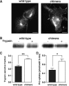
NTT4/XT1 chimeric protein exhibits enhanced plasma membrane expression and transport activity in HEK293T cells. A, heterologously expressed wild-type (left) and chimeric (right) NTT4/XT1 proteins were visualized in transfected HEK293T cells by fluorescence microscopy (scale bar, 10μm). The wild-type protein predominantly localizes to an intracellular population of vesicles. Expression of the chimeric NTT4/XT1 protein in which the carboxyl terminus has been replaced with that of B0AT2/SBAT1 enhances staining of the plasma membrane (arrows). B, immunoblotting of cell extracts with α-HA antibodies shows the relative amount of full-length heterologously expressed wild-type (left) and chimeric (right) NTT4/XT1 proteins before (–) and after (+) 10 min of exposure of suspended cells to trypsin. C, trypsin-sensitive fraction of wild-type and chimeric proteins was quantified as the fractional decrease in the intensity of immunoblotted signal corresponding to the presence of full-length heterologously expressed protein. The trypsin-sensitive fraction is 1.5-fold greater in cells expressing the chimeric protein (** indicates p < 0.01). D, Pro uptake (pmol of Pro/well/5 min) was quantified in HEK293T cells transfected with 0.5 μg of control (i.e. nonfunctional sialin), NTT4/XT-wt, or NTT4/XT1-chi plasmid DNA after a 5-min incubation of cells in assay buffer (120 mm NaGluc, pH 8.5, ** indicates p < 0.01). E, specific NTT4/XT1-mediated Pro uptake (pmol/well/5 min) was quantified by subtracting uptake in cells transfected with control DNA from uptake in cells expressing NTT4/XT1-wt or NTT4/XT1-chi (* indicates p < 0.05).
FIGURE 3.
FIGURE 3.
NTT4/XT1 mediates Na+-dependent, Cl-independent Pro uptake in transfected HEK293T cells. A, Pro uptake was quantified in HEK293T cells co-transfected with 0.25 μg of dynamin K44A plasmid DNA and either 0.5 μg of NTT4/XT1-chi or control (i.e. nonfunctional sialin) plasmid DNA. Analysis of Pro uptake (plotted as pmol/well/5 min) during a 5-min incubation in assay buffer (120 mm NaGluc) demonstrates a 3.2- and 2.9-fold increase over background at pH 7.4 and pH 8.5, respectively. B, Pro uptake into transfected HEK293T cells was quantified after a 5-min incubation of cells in assay buffer, pH 8.5, containing either 120 mm NaCl (left), 120 mm NaGluc (center), or 120 mm NMDG-tartrate (right), demonstrating Cl independence and Na+ dependence of transport. NTT4/XT1-chi-mediated Pro uptake was obtained by subtracting uptake in cells transfected with control DNA from uptake in cells transfected with NTT4/XT1-chi DNA. C, time course of Pro uptake into HEK293T cells transfected with NTT4/XT1-chi or control DNA after incubation of cells in assay buffer (120 mm NaGluc, pH 8.5) for various lengths of time demonstrates greater uptake in the NTT4/XT1-chi-transfected cells at all time points. Plotted data are from duplicate samples of a representative experiment. D, NTT4/XT1-chi-mediated proline uptake was determined by subtracting uptake in control cells from uptake in NTT4/XT1-chi-expressing cells. Uptake is saturated by 20 min. Asterisks in A and B indicate statistical significance (p < 0.01).
FIGURE 4.
FIGURE 4.
NTT4/XT1-mediated Pro uptake is dependent on extracellular [Na+], membrane potential, and pH. A, NTT4/XT1-chi-mediated Pro uptake quantified after a 5-min incubation of transfected HEK293T cells in assay buffer, pH 8.5, containing varying concentrations of Na+ tartrate with ionic substitution of NMDG-tartrate to maintain osmolarity demonstrates a hyperbolic dependence on [Na+]. The difference in uptake between NTT4/XT1-chi-expressing cells and control cells at each extracellular [Na+] was normalized to the subtracted value obtained at 120 mm Na+. B, NTT4/XT1-chi-mediated Pro uptake quantified after a 5-min incubation of transfected HEK293T cells in assay buffer (120 mm NaGluc, pH 8.5) either alone (control) or with the addition of 20 μm valinomycin (val), 25 mm potassium tartrate (K+), or both (val + K+) indicates additive inhibition by valinomycin and potassium. Subtracted values were normalized to assay buffer alone. Conditions with significant differences from control are denoted by single (p < 0.05) or double asterisks (p < 0.01). C, NTT4/XT1-chi-mediated Pro uptake quantified after a 5-min incubation of transfected HEK293T cells in assay buffer (120 mm NaGluc) at pH 5.5, 6.5, 7.2, 8, 8.5, and 9 indicates a strong inhibition by low pH. Subtracted values were normalized to data obtained at pH 8.5. D, NTT4/XT1-chi-mediated Pro uptake was quantified after a 5-min incubation of transfected HEK293T cells in assay buffer (120 mm NaGluc) at pH 8.5, 7.2, and 6.5 in the absence or presence of 10 mm ammonium tartrate. Addition of NH+4 attenuates uptake in the presence of an outwardly directly proton gradient, but it has no effect in the absence of a proton gradient or the presence of an inwardly directed proton gradient. Subtracted values were normalized to data obtained at pH 8.5 in the absence of NH+4. Double asterisks indicate that values are statistically different (p < 0.01).
FIGURE 5.
FIGURE 5.
NTT4/XT1-mediated uptake is differentially inhibited by various amino acids and exhibits substrate-specific kinetic behavior. A, NTT4/XT1-chi-mediated Pro uptake quantified after a 5-min incubation of transfected cells in assay buffer (120 mm NaGluc, pH 8.5) in the absence or presence of 5 mm unlabeled amino acids indicates greatest inhibition by neutral amino acids. Subtracted data were normalized to proline uptake in the absence of unlabeled amino acids. Single (p < 0.05) or double asterisks (p < 0.01) indicate significant differences from uptake in the absence of unlabeled substrate. Standard three letter abbreviations are used for amino acids. l-Pyroglutamic acid, α-methylaminoisobutyric acid, α-aminoisobutyric acid, and γ-aminobutyric acid are indicated by Pyr, MeAIB, AIB, and GABA, respectively. B, measurement of Pro uptake in assay buffer (120 mm NaGluc, pH 8.5) with 0.1 (zero is not shown here) to 10 mm unlabeled Leu, Pro, or Gln demonstrates that Leu and Pro are more effective inhibitors of 3H uptake than Gln. Data are normalized to [3H]Pro uptake observed in the absence of unlabeled substrate. C, analysis of specific NTT4/XT1-chi-mediated uptake of radiolabeled Gln or Pro quantified after a 5-min incubation of transfected cells in assay buffer (120 mm NaGluc, pH 8.5) plus 0–10 mm of the corresponding unlabeled amino acid indicates that both amino acids are transported, but with different Michaelis-Menten kinetics and saturability. Uptake (picomoles of substrate/well/5 min) is plotted as subtracted values for NTT4/XT1-chi-transfected minus control transfected cells and fit with a nonlinear regression. D, summary of B and C. IC50m) represents the concentration of unlabeled substrate that inhibits specific NTT4/XT1-chi-mediated [3H]Pro uptake by 50% as calculated from the linear regression in B. Km and Vmax values are derived from the nonlinear regression of [3H]Pro and [3H]Gln uptake in the presence of increasing concentrations of unlabeled Pro and Gln (C). Km (mm) is the concentration of substrate at which the rate of uptake is half-maximal.
FIGURE 6.
FIGURE 6.
PC12 cells expressing wild-type NTT4/XT1 exhibit Pro uptake activity that is enhanced in the presence of a calcium ionophore. A, differentiated PC12 cells stably expressing HA-tagged wild-type NTT4/XT1 protein were co-immunostained with α-HA (red) and α-synaptobrevin (green) antibodies. An enlarged view of the demarcated region is shown in the bottom panels. Scale bars in both sets of images correspond to 10 μm. Arrows denote puncta along a cellular process that exhibit co-localization of NTT4/XT1 and synaptobrevin. B, uptake of Pro by wild-type PC12 cells and NTT4/XT1-expressing PC12 cells was quantified after a 5-min incubation in assay buffer (120 mm NaCl, pH 8.5). Normalization to uptake by wild-type PC12 cells shows a 2.4-fold increase in Pro transport by NTT4/XT1-expressing cells. C, NTT4/XT1-mediated Pro uptake in PC12 cells was quantified after a 5-min incubation in assay buffer (120 mm NaCl, pH 8.5) in the presence or absence of 2.2 mm CaCl2 either alone (black) or with addition of 0.5 μm ionomycin (white). Uptake is plotted as subtracted values (NTT4/XT1-expressing PC12 cells minus wild-type PC12 cells) normalized to uptake in the presence of Ca2+ alone. Asterisks in B and C denote p < 0.01.

Similar articles

Cited by

References

    1. Ferguson, S. M., Savchenko, V., Apparsundaram, S., Zwick, M., Wright, J., Heilman, C. J., Yi, H., Levey, A. I., and Blakely, R. D. (2003) J. Neurosci. 23 9697–9709 - PMC - PubMed
    1. Edwards, R. H. (2007) Neuron 55 835–858 - PubMed
    1. McKenna, M. C. (2007) J. Neurosci. Res. 85 3347–3358 - PubMed
    1. Chen, N. H., Reith, M. E., and Quick, M. W. (2004) Pfluegers Arch. 447 519–531 - PubMed
    1. Broer, S. (2006) Neurochem. Int. 48 559–567 - PubMed

Publication types

MeSH terms