. 2009 Jan;7(1):33-40.
doi: 10.1158/1541-7786.MCR-08-0369.

Two tumor suppressors, p27Kip1 and patched-1, collaborate to prevent medulloblastoma

Affiliations

Two tumor suppressors, p27Kip1 and patched-1, collaborate to prevent medulloblastoma

Olivier Ayrault et al. Mol Cancer Res. 2009 Jan.

Abstract

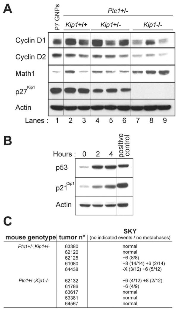
Two cyclin-dependent kinase inhibitors, p18(Ink4c) and p27(Kip1), are required for proper cerebellar development. Loss of either of these proteins conferred a proliferative advantage to granule neuron progenitors, although inactivation of Kip1 exerted a greater effect. Mice heterozygous for Patched-1 (Ptc1+/-) that are either heterozygous or nullizygous for Kip1 developed medulloblastoma rapidly and with high penetrance. All tumors from Ptc1+/-;Kip1+/- or Ptc1+/-;Kip1-/- mice failed to express the wild-type Ptc1 allele, consistent with its role as a canonical "two-hit" tumor suppressor. In contrast, expression of the wild-type p27(Kip1) protein was invariably maintained in medulloblastomas arising in Ptc1+/-;Kip1+/- mice, indicating that Kip1 is haploinsufficient for tumor suppression. Although medulloblastomas occurring in Ptc1+/- mice were histopathologically heterogeneous and contained intermixed regions of both rapidly proliferating and nondividing more differentiated cells, tumors that also lacked Kip1 were uniformly less differentiated, more highly proliferative, and invasive. Molecular analysis showed that the latter medulloblastomas exhibited constitutive activation of the Sonic hedgehog signaling pathway without loss of functional p53. Apart from gains or losses of single chromosomes, with gain of chromosome 6 being the most frequent, no other chromosomal anomalies were identified by spectral karyotyping, and half of the medulloblastomas so examined retained a normal karyotype. In this respect, this mouse medulloblastoma model recapitulates the vast majority of human medulloblastomas that do not sustain TP53 mutations and are not aneuploid.


Figures

FIGURE 1
FIGURE 1
Expression of cell cycle regulators during cerebellar development. A. Expression of proteins (indicated at the left of the panel) was detected by immunoblotting of protein lysates generated from the cerebella of postnatal (P1 to P30) wild type mice. Actin was used as a loading control. B. The bar graphs indicate the percentage of primary GNPs that incorporated BrdU under different culture conditions (panels a, b, and c). GNPs were purified from the cerebella of P10 mice of different genotypes [wild type (wt) (black bar), Ink4c-null (unfilled bar), and Kip1-null (shaded bar)]. GNPs were cultured for one day (panel a) or 3 days (panel b) without Shh, or cultured one day without Shh followed by 2 days with Shh (panel c). BrdU was added 24 hours before the fixation of the cells.
FIGURE 2
FIGURE 2
Kip1 loss increases the incidence of MBs in Ptc1 heterozygous mice. A. Panel a illustrates survival curves for Ptc1+/− mice retaining two wild type Kip1 alleles (+/+: black line, n= 70), or lacking one (+/−: grey dotted line, n= 144) or two (−/− grey line, n= 30) Kip1 alleles. The incidence of MBs and other tumors arising in these cohorts is indicated in the Table in panel b. Mice showing signs of disease without any obvious brain tumors developed hydrocephaly, hemangiosarcomas, intestinal tumors, rhabdomyosarcomas or lymphomas. Not all mice succumbed to tumors. B. Survival curves of Ptc1+/− mice of different Kip1 genotypes with confirmed MB development. Line designations are the same as for panel a; the number of mice in each cohort was 30 for Kip1+/+, 86 for Kip1+/−, and 20 for Kip1−/− genotypes. C. Detection by immunoblotting of the p27Kip1 protein in GNPs purified from a P7 wild type cerebellum and in purified tumor cells from MBs arising in Ptc1+/−;Kip1+/+ and Ptc1+/−;Kip1+/− mice as designated at the top of the panels. Actin was used a loading control. D. Expression of p27Kip1 determined by immunohistochemistry (brown) in cerebellar sections from Ptc1+/− mice of different Kip1 genotypes. All sections were counterstained with hematoxylin (blue). Panels a, b, and c illustrate sections taken from normal cerebellar tissues from Ptc1+/−;Kip1+/+, Ptc1+/−;Kip1−/−, and Ptc1+/−;Kip1+/− mice, respectively. Panels d, e, and f illustrate p27Kip1 expression in MBs arising in Kip1+/+, Kip1−/−, and Kip1+/− mice, respectively. Panel b was photographed at 10X magnification; the unstained region represents the external molecular layer of the adult cerebellum which is practically devoid of cell bodies. All other panels were photographed at 40X magnification. Panels a and c denote cells within the p27Kip1-positive IGL.
FIGURE 3
FIGURE 3
Pathological characteristics of MBs. Tumors from Ptc1+/−;Kip1+/+ mice (panels A–D) were non invasive and confined to the periphery of the cerebellum (A), whereas those arising in Ptc1+/−;Kip1−/− mice (panels E–H) were more invasive and extended into the white matter (E). Interrupted black dashes in E designate the contours of the invaded lobes. Sections were stained with hematoxylin and eosin (A, B, E, F) and by immunohistochemistry with antibodies to Ki-67 (C and G) or to NeuN (D and H) with hematoxylin counterstain. Tumors that retained both Kip1 alleles commonly exhibited a biphasic pattern (B) in which differentiated Ki-67-negative and Neu-N-positive neurons were surrounded by proliferating Ki-67-positive and NeuN-negative cells (serial sections shown in C and D). Kip1−/− MBs were comprised of more uniform sheets of tumor cells (F) that were Ki-67-positive (G) and NeuN-negative (H). Magnifications were 2X (panel A), 40X (panels B, C, D and F) and 4X (panels E, G and H).
FIGURE 4
FIGURE 4
Molecular analysis of GNPs and MBs. A. Immunoblotting of the indicated proteins (left) expressed in primary GNPs purified from a cerebellum of a P7 wild type mouse and from GNP-like tumor cells purified from MBs occurring in Ptc1+/− mice lacking no (+/+), one (+/−) or two (−/−) Kip1 alleles. Actin was used as loading control. B. Accumulation of p53 and p21Cip1 2 and 4 hours after exposure to 15 Gy ionizing radiation in tumor cells purified from MBs from Ptc1+/−;Kip1−/− mice. Zn2+-treated MT-Arf cells were used a positive control. C. Spectral karyotyping (SKY) of MBs from representative tumors arising in Ptc1+/−;Kip1+/− and Ptc1+/−;Kip1−/− animals was used to determine the nature and frequency of chromosomal anomalies.

References

    1. Marino S. Medulloblastoma: developmental mechanisms out of control. Trends Mol Med. 2005;11:17–22. - PubMed
    1. Wang VY, Zoghbi HY. Genetic regulation of cerebellar development. Nat Rev Neurosci. 2001;27:484–91. - PubMed
    1. Ciemerych MA, Kenney AM, Sicinska E, Kalaszczynska I, Bronson RT, Rowitch DH, et al. Development of mice expressing a single D-type cyclin. Genes Dev. 2002;16:3277–89. - PMC - PubMed
    1. Zindy F, Knoepfler PS, Xie S, Sherr CJ, Eisenman RN, Roussel MF. N-Myc and the cyclin-dependent kinase inhibitors p18Ink4c and p27Kip1 coordinately regulate cerebellar development. Proc Natl Acad Sci USA. 2006;103:11579–83. - PMC - PubMed
    1. Sherr C, Roberts JM. CDK inhibitors: positive and negative regulators of G1-phase progression. Genes Dev. 1999;13:1501–12. - PubMed

Publication types

MeSH terms