. 2009 Jan 15;69(2):510-7.
doi: 10.1158/0008-5472.CAN-08-0858.

Identification of nuclear export inhibitors with potent anticancer activity in vivo

Affiliations

Identification of nuclear export inhibitors with potent anticancer activity in vivo

Sarah C Mutka et al. Cancer Res. .

Abstract

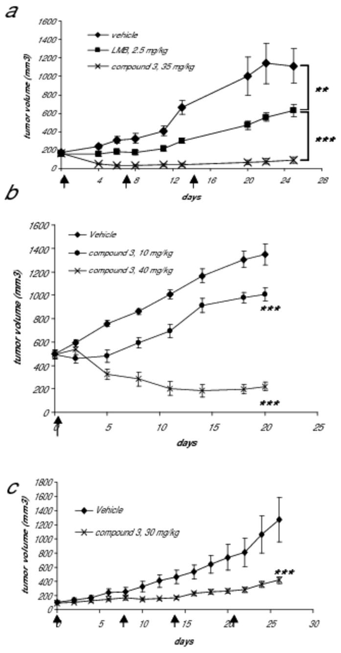
The export protein CRM1 is required for the nuclear export of a wide variety of cancer-related "cargo" proteins including p53, c-Abl, and FOXO-3A. Leptomycin B (LMB) is a highly specific inhibitor of CRM1 with significant in vitro potency but limited in vivo efficacy due to toxicity. We now report a series of semisynthetic LMB derivatives showing substantially improved therapeutic windows. Exposure of cancer cells to these compounds leads to a rapid and prolonged block of nuclear export and apoptosis. In contrast to what is observed in cancer cells, these agents induce cell cycle arrest, but not apoptosis, in normal lung fibroblasts. These new nuclear export inhibitors (NEI) maintain the high potency of LMB, are up to 16-fold better tolerated than LMB in vivo, and show significant efficacy in multiple mouse xenograft models. These NEIs show the potential of CRM1 inhibitors as novel and potent anticancer agents.


Figures

Figure 1
Figure 1. Nuclear export is rapidly blocked by treatment with LMB or compound 3
SiHa (human cervical cancer) cells were treated with 10 nM compound 3 or LMB for the indicated timepoints up to two hours. Cells were formaldehyde fixed and stained for RanBP1 (a biomarker for CRM1 inhibition; red) and Hoescht (blue, to define the nucleus). (a) RanBP1 is localized to the cytoplasm in untreated cells. (b) After drug treatment, nuclear localization of RanBP1 is detected in essentially 100% of the cells. (c) For each timepoint, images were acquired and analyzed using a Cellomics ArrayScan Vti with Molecular Translocation software. By 30 minutes, RanBP1 is localized to the nucleus in the entire population. A minimum of 500 cells per well was analyzed.
Figure 2
Figure 2. Nuclear export block persists after removal of NEI
SiHa (human cervical cancer) cells were treated with 5 nM compound 3 for 1 hour. The drug was removed by washing, and cells were allowed to recover in drug-free media for the times indicated. Cells were formaldehyde fixed and stained for RanBP1 (a biomarker for CRM1 inhibition; red) and Hoescht (blue, to define the nucleus). (a) In untreated cells, RanBP1 is localized to the cytoplasm. After one hour of drug treatment with compound 3, nuclear localization of RanBP1 is detected in essentially 100% of the cells. (b) For each timepoint, images were acquired and analyzed using a Cellomics ArrayScan Vti with Molecular Translocation software. A minimum of 500 cells per well was analyzed, and data shown is the average of two independent experiments. Nuclear export remains blocked in >80% of the cell population 8 hours after the drug is removed and a minority (~25%) of the cells remain at least partially blocked 24 hours after the drug is removed.
Figure 3
Figure 3. NEI treatment induces apoptosis in cancer cells but not in normal lung fibroblasts
(a) SiHa human cervical cancer cells or MRC5 human normal lung fibroblast cells were treated with either vehicle or 10 nM compound 3 for 1h and incubated for an additional 72 hours. Cells were stained with Annexin V-FITC and propidium iodide and analyzed by flow cytometry to determine the Annexin V positive, propidium iodide negative population. Data is representative of multiple experiments. (b) SiHa (cervical cancer cells, black bars), or human normal lung fibroblast cell lines MRC5 (dark gray bars), WI-38 (light gray bars), or IMR-90 (hatched bars) were treated with either vehicle or a high dose (500 nM) of compound 3 or LMB for 1h and incubated for an additional 72 hours. Staurosporine control treatment was at 1 microM for 72 hours. Cells were analyzed for caspase 3/7 induction using the Promega Caspase 3/7 Glo assay. Data is the average of two independent wells and representative of multiple experiments.
Figure 4
Figure 4. Nuclear export inhibition correlates with sustained p53 overexpression and loss of p53 function negatively affects NEI-induced apoptosis
(A) A U20S osteosarcoma cell line containing an NES-RevGFP reporter protein (32) was treated with increasing concentrations of compound 3. After 24 hours of continuous drug treatment, cells were formaldehyde fixed and stained for p53 and with Hoescht dye. For each concentration, images were acquired and a minimum of 500 cells per time point were analyzed using a Cellomics ArrayScan Vti with Compartmental Analysis software (Pittsburg, PA.) In untreated cells, the RevGFP fusion protein localizes predominantly to the cytoplasm but treatment with compound 3 causes accumulation of RevGFP in the nucleus (squares). p53 expression levels (open circles) correlate with the level of nuclear export block. Western blot analysis confirms the upregulation of p53 expression in response to nuclear export inhibition. (b) HCT-116 colon cancer cells were treated with 10 nM compound 3 for one hour followed by a recovery period in drug free media. Cell extracts were made at the indicated times and analyzed by SDS-PAGE for p53 expression levels. GAPDH is shown as a control for protein loading levels. (c). SiHa cells were treated with 50 nM compound 3 in the presence or absence of 10 or 30 μM pifithrin-alpha, an inhibitor of p53. After 72 hours, caspase activation was measured using the Promega Caspase 3/7 Glo assay. As a positive control for apoptosis, cells were treated with 1 μM staurosporine.
Figure 5
Figure 5. Compound 3 inhibits tumor growth in cancer xenograft models
(a) SiHa tumor bearing mice (n=6 each group) were treated i.v. with either vehicle, 2.5 mg/kg LMB, or 35 mg/kg compound 3 using a Q7D schedule for a total of 3 doses (dosing days indicated by arrows). Tumor growth curves of vehicle- or NEI-treated groups are shown. In the compound 3-treated group, 2 mice (~33%) had no detectable tumors after day 6 and remained tumor free at day 25 of the follow up period. **, p<0.01; ***, p<0.001 (based on a 2-way ANOVA test). (b) SiHa xenograft tumors (n=6 each group) were allowed to reach ~500 mm3 before dosing (dosing day indicated by an arrow). A single i.v. bolus dose of compound 3 produced growth inhibition at 10 mg/kg or regression at 40 mg/kg of large SiHa tumors. ***, p<0.001 (based on a 2-way ANOVA test). (c) HCT-116 tumor bearing mice were treated i.v. with either vehicle (n=7) or 30 mg/kg of compound 3 (n=8) using a Q7D schedule for a total of 4 doses (dosing days indicated by arrows). Tumor growth curves are shown. ***, p<0.001 (based on a 2-way ANOVA test).

References

    1. Vousden KH, Woude GF. The ins and outs of p53. Nat Cell Biol. 2000;2:E178–80. - PubMed
    1. Nishi K, Yoshida M, Fujiwara D, Nishikawa M, Horinouchi S, Beppu T. Leptomycin B targets a regulatory cascade of crm1, a fission yeast nuclear protein, involved in control of higher order chromosome structure and gene expression. J Biol Chem. 1994;269:6320–4. - PubMed
    1. Wolff B, Sanglier JJ, Wang Y. Leptomycin B is an inhibitor of nuclear export: inhibition of nucleo-cytoplasmic translocation of the human immunodeficiency virus type 1 (HIV-1) Rev protein and Rev-dependent mRNA. Chem Biol. 1997;4:139–47. - PubMed
    1. Kudo N, Matsumori N, Taoka H, et al. Leptomycin B inactivates CRM1/exportin 1 by covalent modification at a cysteine residue in the central conserved region. Proc Natl Acad Sci U S A. 1999;96:9112–7. - PMC - PubMed
    1. Petosa C, Schoehn G, Askjaer P, et al. Architecture of CRM1/Exportin1 suggests how cooperativity is achieved during formation of a nuclear export complex. Mol Cell. 2004;16:761–75. - PubMed

Publication types

MeSH terms