. 2009 Jan 15;69(2):616-24.
doi: 10.1158/0008-5472.CAN-08-0811.

Melanoma antigen-11 inhibits the hypoxia-inducible factor prolyl hydroxylase 2 and activates hypoxic response

Affiliations

Melanoma antigen-11 inhibits the hypoxia-inducible factor prolyl hydroxylase 2 and activates hypoxic response

Olga Aprelikova et al. Cancer Res. .

Abstract

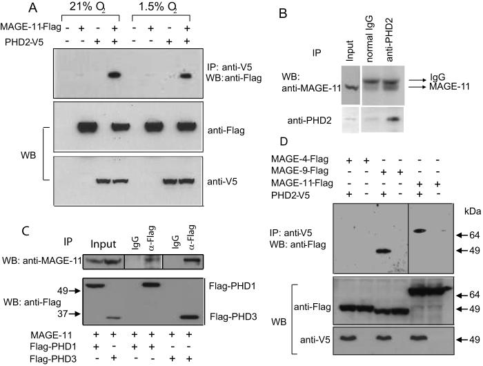
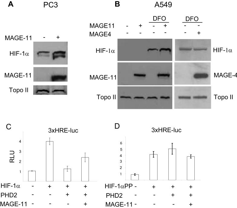
Activation of hypoxia-inducible factors (HIF), responsible for tumor angiogenesis and glycolytic switch, is regulated by reduced oxygen availability. Normally, HIF-alpha proteins are maintained at low levels, controlled by site-specific hydroxylation carried out by HIF prolyl hydroxylases (PHD) and subsequent proteasomal degradation via the von Hippel-Lindau ubiquitin ligase. Using a yeast two-hybrid screen, we identified an interaction between melanoma antigen-11 (MAGE-11) cancer-testis antigen and the major HIF-alpha hydroxylating enzyme PHD2. The interaction was confirmed by a pull-down assay, coimmunoprecipitation, and colocalization in both normoxic and hypoxic conditions. Furthermore, MAGE-9, the closest homologue of MAGE-11, was also found to interact with PHD2. MAGE-11 inhibited PHD activity without affecting protein levels. This inhibition was accompanied by stabilization of ectopic or endogenous HIF-1alpha protein. Knockdown of MAGE-11 by small interfering RNA results in decreased hypoxic induction of HIF-1alpha and its target genes. Inhibition of PHD by MAGE-11, and following activation of HIFs, is a novel tumor-associated HIF regulatory mechanism. This finding provides new insights into the significance of MAGE expression in tumors and may provide valuable tools for therapeutic intervention because of the restricted expression of the MAGE gene family in cancers, but not in normal tissues.


Figures

Figure 1
Figure 1
MAGE-11 interaction with PHD2. (A) Flag-MAGE-11 and V5-PHD2 were cotransfected into 293T cells and two days later treated with 1.5% oxygen or left untreated for 16 hours. Immunoprecipitation was performed with anti-V5 agarose, followed by Western blotting with anti-Flag antibodies. (B) Co-immunoprecipitation of endogenous MAGE-11 and PHD2 from HeLa cell extracts. Protein complexes were immunoprecipitated with anti-PHD2 antibody or normal rabbit IgG, as a negative control, followed by western blotting with anti-MAGE-11 (top panel) or anti-PHD2 antibody (lower panel). (C) MAGE-11 was overexpressed in 293T cells with Flag-PHD1 or Flag-PHD3 and immunoprecipitated with anti-Flag or normal mouse IgG, followed by Western blotting with anti-MAGE-11 antibody. (D) Three different Flag-tagged MAGEA genes were overexpressed in 293T cells alone or in combination with V5-PHD2 and immunoprecipitated with anti-V5 agarose followed by Western blotting with anti-Flag antibody.
Figure 2
Figure 2
PHD2 co-localizes with MAGE-11 in HeLa cells. Confocal microscopy of HeLa cells exposed to 0.5% or 21% oxygen and stained with affinity purified anti-MAGE-11 rabbit antibody and anti-PHD2 mouse antibody followed by secondary anti-rabbit-Alexa 568 (green fluorescence) and anti-mouse-Alexa 486 (red fluorescence) antibody.
Figure 3
Figure 3
MAGE-11 suppresses enzymatic activity of PHD2. (A) V5-PHD2 and increasing amounts of Flag-MAGE-11 were co-expressed in HeLa and Hep3B cells, and 48 hours later, 100 μg of protein were used to hydroxylate HIF-1α-derived 19 amino acid peptide (HIF19), followed by binding with in vitro translated HA-VHL (top panels). The last lanes in both panels show VHL binding to the same peptide that was chemically hydroxylated on Pro-564 and served as a positive control. The numbers under the top panels show the intensity of each HA-VHL band obtained by gel densitometry. Bottom panels show that increase in MAGE-11 protein does not affect PHD2 protein levels. (B) MAGE-4 does not change PHD activity when used instead of MAGE-11. (C) Overexpression of MAGE-11 in the MDA-MB435 cells does not change the levels of endogenous PHD2. UB - unspecific band that serves as a loading control.
Figure 4
Figure 4
MAGE-11 increases the stability and transcriptional activity of HIF-1α. (A) A plasmid containing Gal4 DNA binding domain and HA-tagged HIF-1α ODD (amino acids 496-626) domain (HA-GHO) was co-transfected with pSG5-MAGE-11, and 2 days later, cells were treated with 10 μM MG132 for 4 hours. Each transfection reaction contained equal amounts of Flag-BAP (Bacterial Alkaline Phosphatase) that served as a transfection efficiency control. (B) Shorter exposure of the fragment shown in the top panel of Fig.4A, allowed the separation and quantitation of the two bands. Numbers under the picture show the percentage of unhydroxylated band (GHO) and hydroxylated band (GHO-OH) in cells with and without MAGE-11. (C) Co-expression with PHDs completely (PHD2) or partially (PHD1 and 3) eliminates the ability of MAGE-11 to stabilize GHO protein. MAGE-11 and HA-GHO were co-transfected into 293T cells with or without Flag-tagged PHD1, 2 or 3. (D) MAGE-11, but not MAGE-4, stabilizes full-length HIF-1α protein, but has no effect on PHD-insensitive HIF-1α mutant. pSG5-MAGE-11 was co-transfected with the full-length wild-type HIF-1α or HIF-1α with two proline residues (P402 and P564) mutated to alanine (HIF-1αPP), and the stability of HIF-1α was analyzed with Western blotting. Equal amounts of pCMV-CD20 plasmid were co-transfected in each condition, and served as a transfection efficiency control. The bottom panel in Fig. 4D shows the amounts of HIF-1α protein normalized to CD20.
Figure 5
Figure 5
MAGE-11 increases the protein abundance and transcriptional activity of endogenous HIF-1α. Endogenous HIF-1α protein was analyzed after overexpression of MAGE-11 in PC3 cells (A) or A549 cells treated with DFO at 100 μM for 16 hours (B). (C and D) HIF-1α (C) or HIF-1αPP (D) were co-transfected into 293T cells with or without PHD2 and MAGE-11 and equal amounts of 3xHRE-luciferase and pCMV-RL as an internal control. The experiments were performed in triplicate and repeated twice. The data show the mean values ± s.e.m.
Figure 6
Figure 6
Downregulation of MAGE-11 by siRNA results in impaired HIF-1α induction by hypoxia and inhibition of hypoxia-induced transcription of HIF target genes. (A) The siRNA targeting MAGE-11 was transfected into U2OS or HeLa cells for two consecutive days and in 24 hours samples were collected for Western blotting (top panels) or quantitative RT-PCR analysis (bottom panels). The results are mean values of relative mRNA induction normalized to beta-actin plus standard errors of the mean of at least three experiments. P values were obtained by paired t tests (**, P<0.005; *, P<0.01). (B) Stable expression of shMAGE-11 in MDA-MB435 cells results in downregulation of MAGE-11 mRNA (left panel) and protein (right panel). (C) Expression of shMAGE-11 did not affect mRNA for HIF-1α, but impaired the hypoxic induction of HIF target gene NDRG1. (D) HIF-1α immunoblot analysis of 25 μg of nuclear extract after hypoxia-reoxygenation of shMAGE-11 MDA-MB435 cells. The experiment was repeated three times. The representative experiment is shown.

References

    1. Wang GL, Jiang BH, Rue EA, Semenza GL. Hypoxia-inducible factor 1 is a basic-helix-loop-helix-PAS heterodimer regulated by cellular O2 tension. Proc Natl Acad Sci U S A. 1995;92:5510–4. - PMC - PubMed
    1. Tian H, McKnight SL, Russell DW. Endothelial PAS domain protein 1 (EPAS1), a transcription factor selectively expressed in endothelial cells. Genes Dev. 1997;11:72–82. - PubMed
    1. Jain S, Maltepe E, Lu MM, Simon C, Bradfield CA. Expression of ARNT, ARNT2, HIF1 alpha, HIF2 alpha and Ah receptor mRNAs in the developing mouse. Mech Dev. 1998;73:117–23. - PubMed
    1. Epstein AC, Gleadle JM, McNeill LA, et al. C. elegans EGL-9 and mammalian homologs define a family of dioxygenases that regulate HIF by prolyl hydroxylation. Cell. 2001;107:43–54. - PubMed
    1. Ivan M, Kondo K, Yang H, et al. HIFalpha targeted for VHL-mediated destruction by proline hydroxylation: implications for O2 sensing. Science. 2001;292:464–8. - PubMed

Publication types

MeSH terms

Substances