. 2009 Jan 15;69(2):632-9.
doi: 10.1158/0008-5472.CAN-08-1191.

Helicobacter pylori cytotoxin-associated gene A activates the signal transducer and activator of transcription 3 pathway in vitro and in vivo

Affiliations

Helicobacter pylori cytotoxin-associated gene A activates the signal transducer and activator of transcription 3 pathway in vitro and in vivo

Dana M Bronte-Tinkew et al. Cancer Res. .

Erratum in

  • Cancer Res. 2013 Jul 1;73(13):4170

Abstract

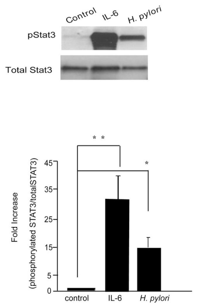
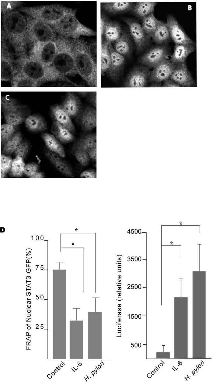
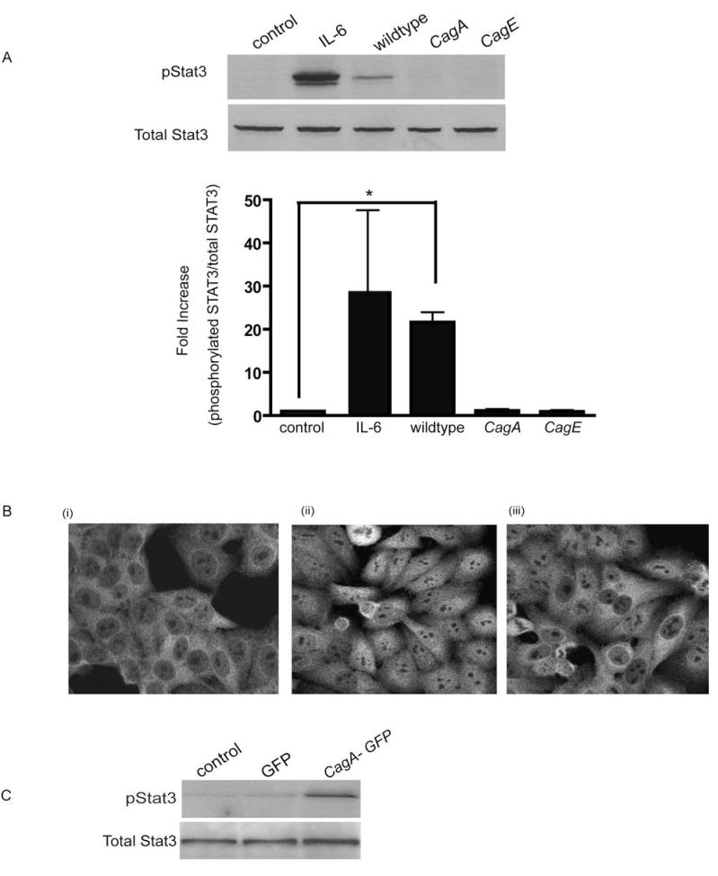
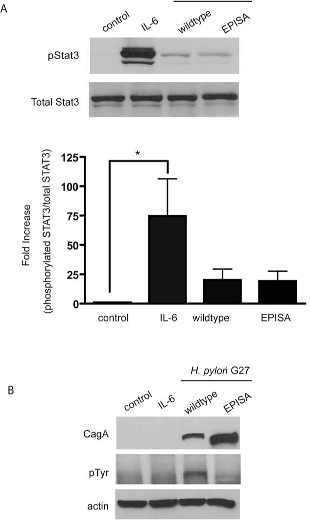
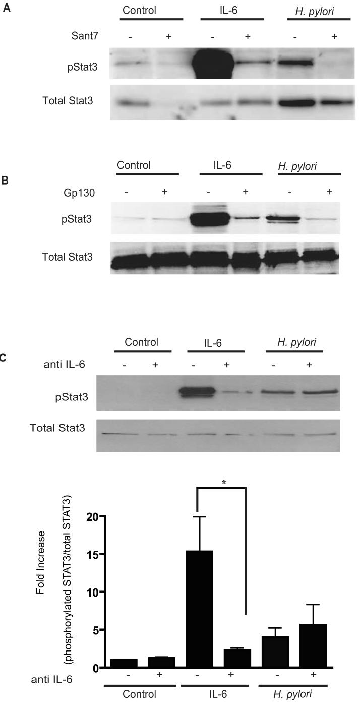
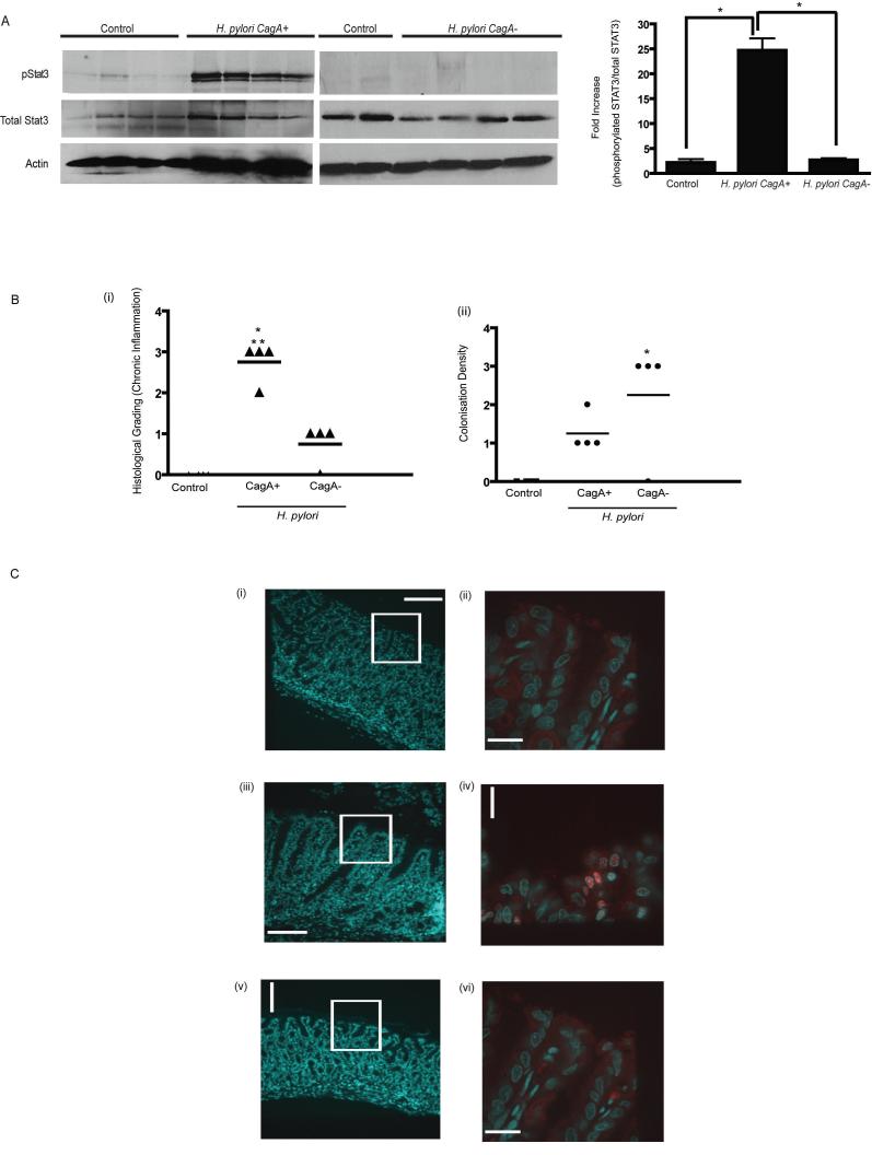
Persistent infection with Helicobacter pylori confers an increased risk for the development of gastric cancer. However, the exact mechanisms whereby this bacterium causes carcinogenesis have not been completely elucidated. Recent evidence indicates that aberrant activation of the signal transducers and activators of transcription 3 (STAT3) signaling pathway may play a role in gastric carcinogenesis. Therefore, we hypothesized that H. pylori infection modulates STAT3 signaling, favoring gastric cancer development. In epithelial cells infected with H. pylori, STAT3 was activated, as assessed by immunoblotting for phosphorylated STAT3, immunofluorescence of translocated STAT3, fluorescence recovery after photobleaching, and luciferase activation in transfected cells. Activation was dependent on translocation but not phosphorylation of cytotoxin-associated gene A (CagA) in host cells. Activation seemed to be receptor-mediated because preincubation of cells with the interleukin-6 (IL-6) receptor superantagonist sant7 or inhibition of gp130 by a monoclonal antibody prevented H. pylori-mediated STAT3 activation. However, activation was not related to autocrine activation by IL-6 or IL-11. CagA+ wild-type H. pylori, but not the noncarcinogenic cagA- mutant, activated STAT3 in gastric epithelial cells in vivo in the gerbil model of H. pylori-mediated gastric carcinogenesis. Collectively, these results indicate that H. pylori CagA activates the STAT3 signaling pathway in vitro and in vivo, providing a potential mechanism by which chronic H. pylori infection promotes the development of gastric cancer.


Figures

Figure 1
Figure 1. H. pylori promotes phosphorylation of STAT3 pathway
Lysates obtained from untreated HEp-2 cells, cells incubated with IL-6 (100ng/ml, 30mins), or cells infected with H. pylori 60190 (wt) (MOI of 100:1) for 4hrs were used for immunoblotting to detect changes in phosphorylated and native STAT3. The graph represents densitometric analysis of the bands obtained for each protein signal normalized to native STAT3. Fold increase given as a ratio of control pSTAT3/STAT3. Results expressed as mean +/- S.E. (** p < 0.01 and * p < 0.05 using the Student’s t test, n=8)
Figure 2
Figure 2. H. pylori induces STAT3 nuclear translocation and transcriptional activation in a CagA-dependent manner
(A-C) HEp-2 cells were treated with culture media (A), IL-6 for 30mins (B) or infected with H. pylori strain 7.13 (C) for 4hrs. Fixed cells were stained with anti-STAT3 and donkey anti-mouse Cy3 conjugated secondary antibodies. Confocal images were taken of representative cells for each treatment. (D) Whole nuclei of uninfected, IL-6-treated and H. pylori infected STAT3-GFP transfected cells were bleached and the percentage recovery of fluorescence was measured relative to the initial values. Six cells for each group were analyzed. (* p <0. 05 using the Student’s t test, n=4). HEp-2 cells were transfected with luciferase reporter constructs containing three repeat STAT3 DNA binding domains, p950m4 or control pSTAT3 plasmids. Transfected cells were treated with IL-6 or infected with H. pylori strain 60190 for 48hrs and luciferase activity was measured and normalised against renilla luciferase activity. (* p <0.05 using the Student’s t test, n=3).
Figure 3
Figure 3. H. pylori-mediated STAT3 activation is CagA dependent
(A) HEp-2 cells were infected with either H. pylori strain 60190, its isogenic cagA mutant or the type IV secretion system mutant cagE (MOI of 100:1 for 4 hrs). Whole cell lysates were collected and probed for STAT3 activation. Untreated and IL-6 treated cells (50ng/ml) served as controls. Graph depicts the densitometric analysis for pSTAT3 bands relative to the native STAT3 protein (* p < 0.05 using the Student’s t test, n=4). (B) (i) Untreated control (ii) H. pylori wildtype or (iii) cagA mutant infected HEp-2 cells were immunolablled for STAT3 and images obtained using confocal micrscopy. (C) HEp-2 cells were transfected with untagged GFP or CagA-GFP for 20hrs, cell lysates collected and probed for phosphoSTAT3.
Figure 4
Figure 4. CagA-dependent STAT3 activation does not require tyrosine phosphorylation of CagA
(A) Cells were infected with strain G27 or its cagA EPISA mutant. Cell lysates were probed for phosphoSTAT3. Graph depicts the density of pSTAT3 protein bands normalized to native STAT3 detected in each treatment (* p < 0.05 using the Student’s t test, n=6). (B) Lysates initially probed for pSTAT3 were subsequently analyzed by immunoblotting using a tyrosine phosphorylation specific antibody to detect the presence of tyrosine phosphorylated CagA.
Figure 5
Figure 5. H. pylori-mediated STAT3 activation requires the IL-6R and the gp130 receptor but is independent of the IL-6 cytokine
(A) Cells were pre-incubated with the IL-6 receptor super-antagonist Sant7 prior to incubation with IL-6 (50ng/ml, 30mins) or infection with strain 60190. Lysates were probed for pSTAT3 and native STAT3 protein. Representative of three independent experiments. (B) An anti-gp130 blocking Ab was incubated with cells prior to either H. pylori infection or incubation with IL-6, cell lysates obtained and immunoblotted for pSTAT3. Representative of three independent experiments. (C) HEp-2 cells were treated with neutralizing IL-6 Ab followed by infection with strain 60190. Whole cell lysates were collected and probed for pSTAT3 via Western Blotting Densitometric analysis showing the ratio of pSTAT3 normalized to native STAT3 was performed and represented in the graph (* p <0.05 using Student’s t test, n=4).
Figure 6
Figure 6. H. pylori carcinogenic strain 7.13 induces STAT3 activation in vivo in a CagA dependent manner
(A) Stomach sections from Mongolian Gerbils sham infected (n=4) or infected with H. pylori strain 7.13 (n=4) or its isogenic cagA- mutant (n=4) for 12 weeks were collected and homogenized to obtain protein lysates. Lysates were probed for pSTAT3, STAT3 and actin. Densitomtric analysis was performed and depicted in graph (* p<0.05 using Student’s t test) (Bi) Severity of Inflammation within the gastric antrum of animals infected with carcinogenic H. pylori strain 7.13. Chronic antral gastritis was scored from numbers 1 to 3 (14) in control, CagA+ and cagA- infected gerbil stomachs and mean values represented on scatter plots. (* p <0.05 for infected gerbils compared to control sham infected animals, **p< 0.05 for H. pylori CagA+ infected gerbils compared with H. pylori cagA- animals). (Bii) Colonization density was quantitated using a standardized grading system (14) and data presented in scatter plot (p< 0.05 for control animals compared to infected gerbils (* p <0.05 for infected gerbils compared to control sham infected animals) (C) Immunohistochemistry showing DAPI and STAT3 staining of cells within the antral mucosa. Panels on the left show DAPI stained sections from (i) control (iii) H. pylori CagA+ and (v) H. pylori cagA- infected stomachs (x 10 magnification). White boxes highlight the corresponding regions magnified in panels (ii), (iv) and (vi) (X 63 magnification) showing anti-pSTAT3 staining (red), and DAPI (blue) co-staining. Spinning Disk Microscopy was utilized to obtain images represented.

References

    1. Peek RM, Jr, Crabtree JE. Helicobacter infection and gastric neoplasia. J Pathol. 2006;208:233–48. - PubMed
    1. Kusters JG, van Vliet AH, Kuipers EJ. Pathogenesis of Helicobacter pylori infection. Clin Microbiol Rev. 2006;3:449–90. - PMC - PubMed
    1. Hatakeyama M. Oncogenic mechanisms of the Helicobacter pylori CagA protein. Nat Rev Cancer. 2004;4:688–94. - PubMed
    1. Mimuro H, Suzuki T, Tanaka J, Asahi M, Haas R, Sasakawa C. Grb2 is a key mediator of Helicobacter pylori CagA protein activities. Mol Cell. 2000;10:745–55. - PubMed
    1. Churin Y, Al Ghoul L, Kepp O, Meyer TF, Birchmeier W, Naumann MJ. Helicobacter pylori CagA protein targets the c-Met receptor and enhances the motogenic response. Cell Biol. 2003;161:249–55. - PMC - PubMed

Publication types

MeSH terms