. 2009 Apr 1;82(1):21-9.
doi: 10.1093/cvr/cvp015. Epub 2009 Jan 15.

MicroRNA expression in response to murine myocardial infarction: miR-21 regulates fibroblast metalloprotease-2 via phosphatase and tensin homologue

Affiliations

MicroRNA expression in response to murine myocardial infarction: miR-21 regulates fibroblast metalloprotease-2 via phosphatase and tensin homologue

Sashwati Roy et al. Cardiovasc Res. .

Abstract

Aims: MicroRNAs (miRNAs) are small non-coding RNAs that regulate gene expression at the post-transcriptional level by either degradation or translational repression of a target mRNA. Encoded in the genome of most eukaryotes, miRNAs have been proposed to regulate specifically up to 90% of human genes through a process known as miRNA-guided RNA silencing. For the first time, we sought to test how myocardial ischaemia-reperfusion (IR) changes miR expression.

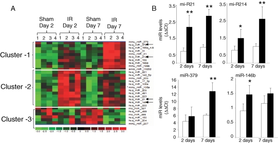
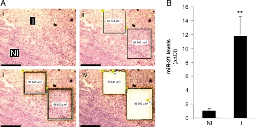
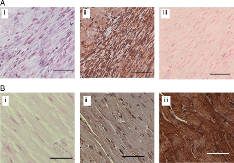
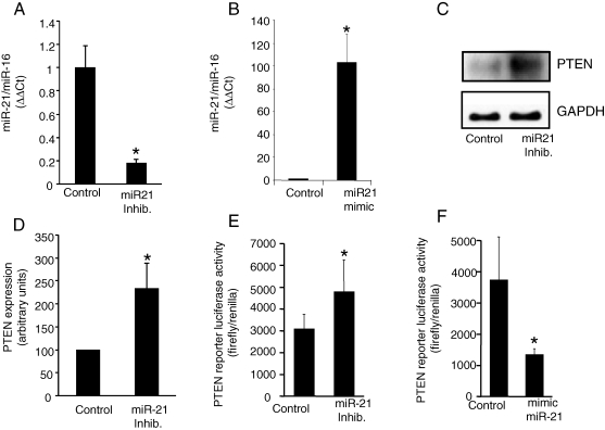
Methods and results: Following 2 and 7 h of IR or sham operation, myocardial tissue was collected and subjected to miRNA expression profiling and quantification using a Bioarray system that screens for human-, mice-, rat-, and Ambi-miR. Data mining and differential analyses resulted in 13 miRs that were up-regulated on day 2, 9 miRs that were up-regulated on day 7, and 6 miRs that were down-regulated on day 7 post-IR. Results randomly selected from expression profiling were validated using real-time PCR. Tissue elements laser-captured from the infarct site showed marked induction of miR-21. In situ hybridization studies using locked nucleic acid miR-21-specific probe identified that IR-inducible miR-21 was specifically localized in the infarct region of the IR heart. Immunohistochemistry data show that cardiac fibroblasts (CFs) are the major cell type in the infarct zone. Studies with isolated CFs demonstrated that phosphatase and tensin homologue (PTEN) is a direct target of miR-21. Modulation of miR-21 regulated expression of matrix metalloprotease-2 (MMP-2) via a PTEN pathway. Finally, we noted a marked decrease in PTEN expression in the infarct zone. This decrease was associated with increased MMP-2 expression in the infarct area.

Conclusion: This work constitutes the first report describing changes in miR expression in response to IR in the mouse heart, showing that miR-21 regulates MMP-2 expression in CFs of the infarct zone via a PTEN pathway.


Figures

Figure 1
Figure 1
miR expression profiling in hearts subjected to ischaemia–reperfusion. (A) Specific clusters of miR showing an increase or decrease in expression following ischaemia (30 min) and reperfusion (2 or 7 days). miRs that were found significantly up- or down-regulated in all replicates following ischaemia–reperfusion (IR) compared with sham-operated samples were selected. These select candidates were subjected to hierarchical clustering to identify clusters of miRs that were induced or down-regulated by ischaemia–reperfusion following 2 or 7 days post-ischaemia–reperfusion. (i) cluster of miR up-regulated specifically 7 days post-ischaemia–reperfusion; (ii) cluster of miR up-regulated 2 days post-ischaemia–reperfusion; (iii) cluster of miR down-regulated specifically 7 days post-ischaemia–reperfusion. Data shown are from four (1–4) individual animals. As is the standard, red to green gradation in colour represents higher to lower expression signals. Annotations of miR in each of the cluster are presented in Table 1. (B) Expression levels of select miR identified using the profiling assay were independently determined using real-time PCR. Real-time PCR data were normalized to miR-16. We chose miR-16 as the normalizing control because the expression of this miR remained unchanged between sham and ischaemia–reperfusion groups. Those marked with a black arrow were verified using quantitative assays. Data represent mean ± SD (n = 4). *P < 0.05 and **P < 0.01 compared with the corresponding sham samples.
Figure 2
Figure 2
Induction of miR-21 in the infarct region of ischaemia-reperfused murine heart: a laser-capture microdissection based approach. C57BL/6 mice were subjected to ischaemia (30 min) and reperfusion (IR). Heart samples were collected 7 days post-IR. (A) Frozen sections (10 µm) from collected heart samples were stained with haematoxylin QS to histologically define the infarct (I) and control or non-infarct (NI) regions. These regions were subsequently cut and captured using laser-capture microdissection for miRNA expression analysis. (i) Representative images of heart tissue showing infarct (I) and control or non-infarct (NI) areas following laser-capture microdissection compatible staining with haematoxylin QS; (ii) the identified area is marked; and (iii) laser-assisted cutting and separation of the identified area for catapulting; (iv) the section after the marked area has been catapulted; Scale bar = 150 µm. (B) The laser-captured tissue elements from I and NI regions were used for quantification of miR-21 expression using real-time PCR. Data represent mean ± SD (n = 4). **P < 0.01 compared with control (NI) tissue.
Figure 3
Figure 3
Localization of miR-21 in ischaemia-reperfused mouse heart regions. Representative image showing localization of miR-21 in a section of a mouse heart subjected to ischaemia for 30 min and reperfusion 7 days. Localization of miR-21 in heart sections was achieved using in situ hybridization analysis with an LNA miR-21-specific probe. (A) miR-21 signal (blue) is evident in the infarct region (i). Fibroblast represented the major cell type in this region as identified using immunohistochemical analysis with an anti-vimentin antibody (ii, brown) The miR-21 signal [as in (i)] was not evident in the serial section when the scrambled LNA miRNA probe was utilized (iii). (B) Representative images from the non-infract region showing no or weak signal (blue) for miR-21 (i). The distribution of fibroblasts and myocytes in this non-infarct (control) region was shown using anti-vimentin (ii, brown, fibroblasts), or anti-muscle actin (iii brown, myocytes) antibody immunohistochemical staining. Nuclear red fast (ISH) and haematoxylin (nuclear, blue, immunohistochemical) were used as counterstains. Scale bar=50 µm. Additional higher magnification images of the infarct region for the ISH and immunohistochemical stains are also presented in Supplementary material online, Figure S3.
Figure 4
Figure 4
Direct regulation of phosphatase and tensin homologue in cardiac fibroblasts by miR-21. After isolation, cardiac fibroblasts were cultured for 5 days. The cells were transfected with miRIDIAN mmu-miR-21 inhibitor or miRIDIAN mmu-miR-21 mimic to down-regulate or increase miR-21 levels. (A and B) miR-21 expression levels in cardiac fibroblasts 72 h post-transfection were determined using real-time PCR. miRIDIAN miRNA inhibitor/mimic negative controls were used for control transfections. Mean ± SD (n = 4), *P < 0.01 compared with control-transfected cardiac fibroblasts. (C) Phosphatase and tensin homologue protein levels in cardiac fibroblasts following knockdown of miR-21 in cardiac fibroblasts. (D) Quantification of phosphatase and tensin homologue level was performed using densitometry. Data were normalized to GAPDH. Data shown represent mean ± SD (n = 3). *P < 0.01 compared with control-transfected cells. (E and F) To demonstrate that phosphatase and tensin homologue is a direct target for miR-21 in cardiac fibroblasts, the cells were transfected with a pGL3-PTEN-3′-UTR firefly luciferase expression construct and co-transfected with pRL-TK Renilla luciferase expression construct along with either miR-21 inhibitor or mimic. An increase in relative firefly luciferase activity in the presence of miR-21 inhibitor or a decrease in the presence of miR-21 mimic indicates that the 3′-UTR of phosphatase and tensin homologue contains a target that is modulated by miR-21. Data represent mean ± SD (n = 3). *P < 0.05 compared with control-transfected cells.
Figure 5
Figure 5
miR-21 regulates MMP-2 expression in cardiac fibroblasts via a PTEN-AKT-phosphorylation-dependent pathway. After isolation, cardiac fibroblasts were cultured for 5 days. The cells were transfected with phosphatase and tensin homologue siRNA. (A) Western blots of AKT phosphorylation and phosphatase and tensin homologue expression in cardiac fibroblasts following transfection of the cells with phosphatase and tensin homologue or control siRNA. β-actin was used for data normalization. (B and C) Quantification of p-AKT and phosphatase and tensin homologue levels were performed using densitometry. Data were normalized to β-actin. Data represent mean ± SD (n = 3). *P < 0.05 compared with control siRNA-transfected cells. (D) MMP-2 expression in cardiac fibroblasts transfected with miR-21 mimic to increase miR-21 levels. MMP-2 expression in cardiac fibroblasts was measured using real-time PCR. Data represent mean ± SD (n = 3). *P < 0.05 compared with mimic control-transfected cells. (E) MMP-2 expression in cardiac fibroblasts transfected either with miR-21 mimic or co-transfected with phosphatase and tensin homologue siRNA. Data represent mean ± SD (n = 3). *P < 0.05 compared with mimic control-transfected cells. #P < 0.05 compared with miR-21 mimic + control siRNA-transfected cells.
Figure 6
Figure 6
Decreased phosphatase and tensin homologue and increased MMP-2 expression in the infarct region of heart. Mice were subjected to left anterior descending coronary artery ligation for 30 min followed by reperfusion (IR). (A) Representative images showing phosphatase and tensin homologue expression in a section of mouse heart subjected to 7 days post-IR. Localization of phosphatase and tensin homologue (green) in heart sections was achieved using anti-phosphatase and tensin homologue antibody. The sections were counterstained with DAPI (nuclear, blue). Myocytes were stained using anti-alpha-sarcomeric actin antibody (red). (i) Representative image of phosphatase and tensin homologue (green) and DAPI (blue), infarct (I) or non-infarct (NI) regions are indicated; (ii) alpha-sarcomeric actin (red) and DAPI (blue) image. Scale bar = 50 µm. (B) Quantification of phosphatase and tensin homologue signal in infarct (I) vs. non-infarct (NI) regions. Data represent mean ± SD (n = 3). *P < 0.05 compared with NI region. (C) Histologically defined infarct (I) and non-infarct (NI) regions were captured using laser-capture microdissection as described in Figure 2. The laser-captured tissue elements from infarct and non-infarct regions were used for quantification of MMP-2 expression using real-time PCR. Data represent mean ± SD (n = 4). **P < 0.01 compared with control (NI) tissue.

Comment in

Similar articles

Cited by

References

    1. Perron MP, Provost P. Protein interactions and complexes in human microRNA biogenesis and function. Front Biosci. 2008;13:2537–2547. - PMC - PubMed
    1. Rajasingh J, Bord E, Hamada H, Lambers E, Qin G, Losordo DW, et al. STAT3-dependent mouse embryonic stem cell differentiation into cardiomyocytes: analysis of molecular signaling and therapeutic efficacy of cardiomyocyte precommitted mES transplantation in a mouse model of myocardial infarction. Circ Res. 2007;101:910–918. - PubMed
    1. Shilo S, Roy S, Khanna S, Sen CK. Evidence for the involvement of miRNA in redox regulated angiogenic response of human microvascular endothelial cells. Arterioscler Thromb Vasc Biol. 2008;28:471–477. - PubMed
    1. Roy S, Khanna S, Bickerstaff AA, Subramanian SV, Atalay M, Bierl M, et al. Oxygen sensing by primary cardiac fibroblasts: a key role of p21(Waf1/Cip1/Sdi1) Circ Res. 2003;92:264–271. - PubMed
    1. Roy S, Khanna S, Rink T, Radtke J, Williams WT, Biswas S, et al. p21waf1/cip1/sdi1 as a central regulator of inducible smooth muscle actin expression and differentiation of cardiac fibroblasts to myofibroblasts. Mol Biol Cell. 2007;18:4837–4846. - PMC - PubMed

Publication types

MeSH terms