. 2009 May;150(5):2491-6.
doi: 10.1210/en.2008-1616. Epub 2009 Jan 15.

Importance of monocarboxylate transporter 8 for the blood-brain barrier-dependent availability of 3,5,3'-triiodo-L-thyronine

Affiliations

Importance of monocarboxylate transporter 8 for the blood-brain barrier-dependent availability of 3,5,3'-triiodo-L-thyronine

Ainhoa Ceballos et al. Endocrinology. 2009 May.

Abstract

Mutations of the gene expressing plasma membrane transporter for thyroid hormones MCT8 (SLC16A2) in humans lead to altered thyroid hormone levels and a severe neurodevelopmental disorder. Genetically engineered defect of the Mct8 gene in mice leads to similar thyroid hormone abnormalities but no obvious impairment of brain development or function. In this work we studied the relative role of the blood-brain barrier and the neuronal plasma cell membrane in the restricted access of T(3) to the target neurons. To this end we compared the effects of low doses of T(4) and T(3) on cerebellar structure and gene expression in wild-type (Wt) and Mct8 null male mice [Mct8-/y, knockout (KO)] made hypothyroid during the neonatal period. We found that compared with Wt animals, T(4) was considerably more potent than T(3) in the Mct8KO mice, indicating a restricted access of T(3), but not T(4), to neurons after systemic administration in vivo. In contrast, T(3) action in cultured cerebellar neurons was similar in Wt cells as in Mct8KO cells. The results suggest that the main restriction for T(3) entry into the neural target cells of the mouse deficient in Mct8 is at the blood-brain barrier.


Figures

Figure 1
Figure 1
Effects of hypothyroidism (Hypo) and thyroid hormone treatment on the structure of the cerebellar cortex of Wt and KO mice. The figure shows photomicrographs of lobule 7 from toloudine-stained sagittal slices of P21 mice. Igl, Internal granular layer; Mol, molecular layer. The arrows show the external germinal layer. Scale bar, 100 μm.
Figure 2
Figure 2
Influence of Mct8 deficit on the response to T4 and T3. Wt and KO mice were made hypothyroid (Hypo) from the late fetal period up to P21, and treated with either vehicle or T4 or T3 for 5 d before the animals were killed. Data are means ± sd. Hr and RC3/Nrgn expression was analyzed by real-time PCR in the cerebellum and striatum, respectively. The data were analyzed by two-way ANOVA and the Bonferroni posttest to compare the data from each KO condition with the corresponding Wt. **, P < 0.01; ***, P < 0.001. For Hr expression, there was a significant effect of genotype (F1,41 = 116.01, P < 0.0001) and thyroidal status (F3,41 = 37.84, P < 0.0001), with a significant interaction (F3,41 = 3.88, P = 0.0156). Also for Nrgn, there was a significant effect of genotype (F1,35 = 24.68, P < 0.0001) and thyroidal status (F3,35 = 41.16, P < 0.0001), with a significant interaction (F3,35 = 10.30, P < 0.0001). The lower panel shows Gpd2 expression in the liver. There was a significant effect of thyroidal status (F3,41 = 23.67, P < 0.0001) but not genotype (F3,41 = 1.065, P = 0.308), with a significant interaction (F3,41 = 3.936, P < 0.0148). NS, Not significant.
Figure 3
Figure 3
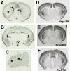
35S in situ hybridization for Mct8 (A–C), Nrgn (D and E) and Dio2 (F) mRNAs. The slices are from coronal sections at the level of the caudate (A, B, and D–F) and sagittal section of the cerebellum (C). All slices are from P21 Wt mice except for E, which shows the typical Nrgn expression in a P21 KO mouse. The arrows in A–C show heavy Mct8 expression in the choroid plexus. The asterisks show the caudate nucleus in A, with low hybridization signal, and the faint but detectable hybridization in the cerebellar cortex in C. 3V, Third ventricle.
Figure 4
Figure 4
Hr expression in primary cultures of granular cells from Wt or Mct8-deficient mice, as a function of T3 added to the cultures. Differences in Hr expression between the cells without T3 added and the 0.2 nm T3 concentration were P < 0.001 (a). Differences between the Wt and KO cells at each T3 concentration, by two-way ANOVA were not significant, except for the 0.25 nm T3, with P < 0.05 (b).

Similar articles

Cited by

References

    1. Dumitrescu AM, Liao XH, Best TB, Brockmann K, Refetoff S 2004 A novel syndrome combining thyroid and neurological abnormalities is associated with mutations in a monocarboxylate transporter gene. Am J Hum Genet 74:168–175 - PMC - PubMed
    1. Friesema EC, Grueters A, Biebermann H, Krude H, von Moers A, Reeser M, Barrett TG, Mancilla EE, Svensson J, Kester MH, Kuiper GG, Balkassmi S, Uitterlinden AG, Koehrle J, Rodien P, Halestrap AP, Visser TJ 2004 Association between mutations in a thyroid hormone transporter and severe X-linked psychomotor retardation. Lancet 364:1435–1437 - PubMed
    1. Refetoff S, Dumitrescu AM 2007 Syndromes of reduced sensitivity to thyroid hormone: genetic defects in hormone receptors, cell transporters and deiodination. Best Pract Res Clin Endocrinol Metab 21:277–305 - PubMed
    1. Grüters A 2007 Thyroid hormone transporter defects. Endocr Dev 10:118–126 - PubMed
    1. Visser WE, Friesema EC, Jansen J, Visser TJ 2008 Thyroid hormone transport in and out of cells. Trends Endocrinol Metab 19:50–56 - PubMed