. 2009 Jan 15;15(2):476-84.
doi: 10.1158/1078-0432.CCR-08-0930.

Mitochondrial mutations contribute to HIF1alpha accumulation via increased reactive oxygen species and up-regulated pyruvate dehydrogenease kinase 2 in head and neck squamous cell carcinoma

Affiliations

Mitochondrial mutations contribute to HIF1alpha accumulation via increased reactive oxygen species and up-regulated pyruvate dehydrogenease kinase 2 in head and neck squamous cell carcinoma

Wenyue Sun et al. Clin Cancer Res. .

Abstract

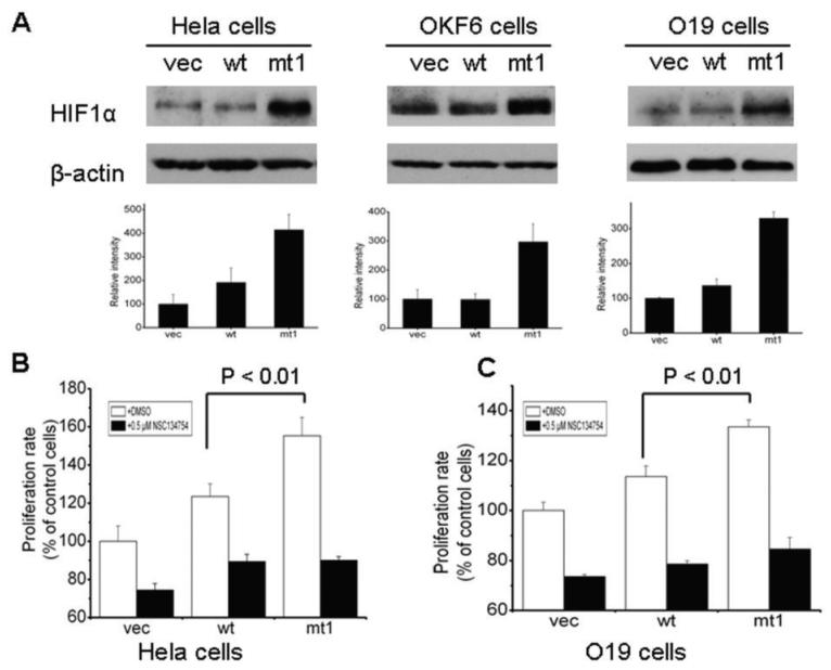
Purpose: Mitochondrial mutations have been identified in head and neck squamous cell carcinoma (HNSCC), but the pathways by which phenotypic effects of these mutations are exerted remain unclear. Previously, we found that mitochondrial ND2 mutations in primary HNSCC increased reactive oxygen species (ROS) and conferred an aerobic, glycolytic phenotype with HIF1alpha accumulation and increased cell growth. The purpose of the present study was to examine the pathways relating these alterations.

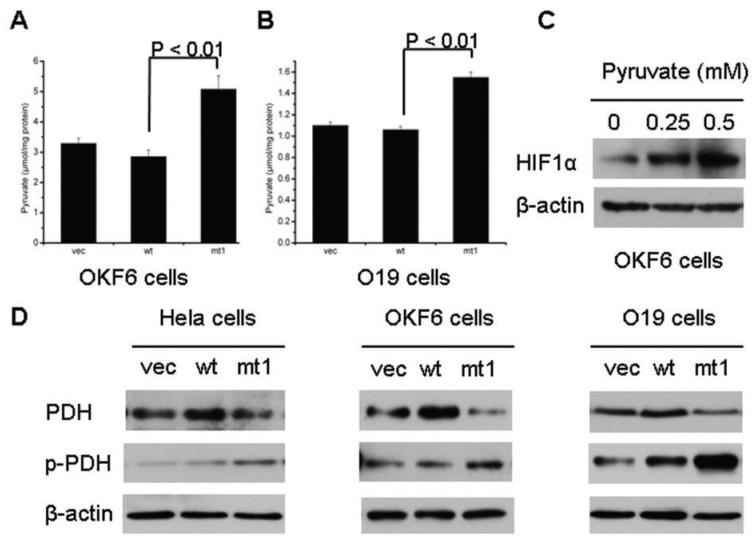
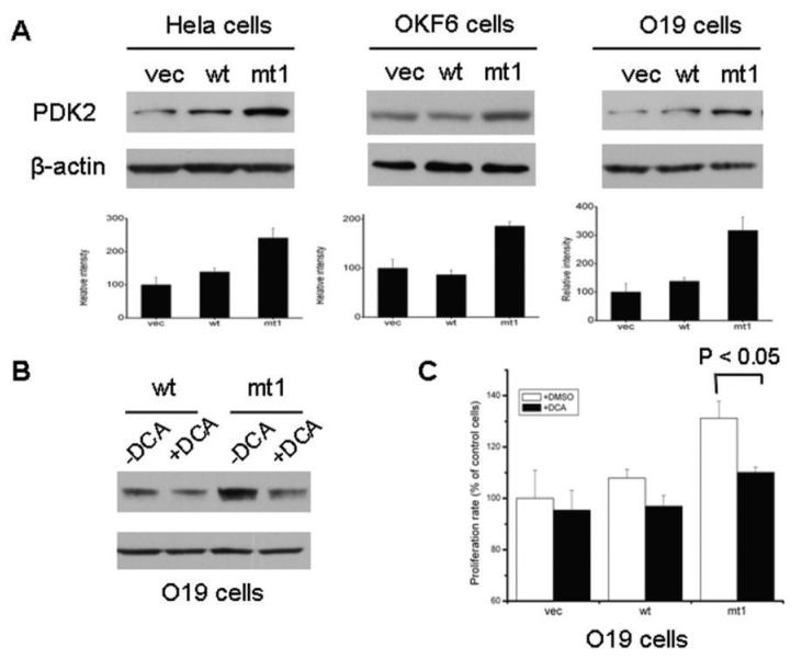
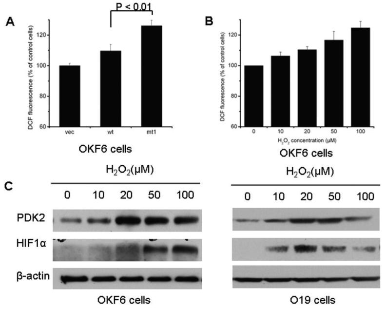
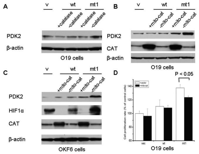
Experimental design: Mitochondrial mutant and wild-type ND2 constructs were transfected into oral keratinocyte immortal cell line OKF6 and head and neck cancer cell line JHU-O19 and established transfectants. The protein levels of HIF1alpha, pyruvate dehydrogenease (PDH), phosphorylated PDH, and pyruvate dehydrogenease kinase 2 (PDK2), together with ROS generation, were compared between the mutant and the wild type. Meanwhile, the effects of small molecule inhibitors targeting PDK2 and mitochondria-targeted catalase were evaluated on the ND2 mutant transfectants.

Results: We determined that ND2 mutant down-regulated PDH expression via up-regulated PDK2, with an increase in phosphorylated PDH. Inhibition of PDK2 with dichloroacetate decreased HIF1alpha accumulation and reduced cell growth. Extracellular treatment with hydrogen peroxide, a ROS mimic, increased PDK2 expression and HIF1alpha expression, and introduction of mitochondria-targeted catalase decreased mitochondrial mutation-mediated PDK2 and HIF1alpha expression and suppressed cell growth.

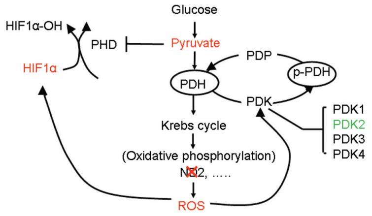
Conclusions: Our findings suggest that mitochondrial ND2 mutation contributes to HIF1alpha accumulation via increased ROS production, up-regulation of PDK2, attenuating PDH activity, thereby increasing pyruvate, resulting in HIF1alpha stabilization. This may provide insight into a potential mechanism, by which mitochondrial mutations contribute to HNSCC development.


Figures

Figure 1
Figure 1
Mitochondrial ND2 mutant promoted head and neck cancer cell growth through HIF1α accumulation. A, the protein levels of HIF1α were determined 4 h following switching from culture medium to Krebs-Henseleit buffer in Hela, OKF6, and O19 cells. Immunoblots were quantified via densitometry and ImageJ software. HIF1α was overexpressed by 3.0 ± 0.9-fold, 4.0 ± 1.7-fold, and 2.5 ± 0.4-fold in the three cells transfected mitochondrial ND2 mutant versus their corresponding wild type (n = 3, P < 0.05). Representative images from three independent experiments were shown. B, the growth of Hela cells stably transfected with ND2 mutant, was inhibited after treatment with NSC134754, a HIF1α small inhibitor. Cell growth was estimated by using a sulforhodamine B assay, after exposure for 48 h to 0.5 μM of NSC134754. C, the growth of O19 cells with ND2 mutant, was inhibited after treatment with NSC134754. Cell growth was estimated by staining with sulforhodamine B, after exposure for 48 h to 0.5 μM of NSC134754. Data are mean ± SD values from three independent experiments. Student’s t test showed significance between mutant and wild type (P < 0.01) in the absence of NSC134754 whereas no significance in the presence of NSC134754. Vec, vector alone; wt, wild-type; mt1, mutant.
Figure 2
Figure 2
Mitochondrial ND2 mutant decreased PDH, increased phospho-PDH, and thereby increased pyruvate production, resulting in increased HIF1α accumulation. A and B, the production of pyruvate in the culture buffer was measured in OKF6 cells and O19 cells. Twenty-four hours after transiently transfection with ND2 mutant and wild type construct, the cells were allowed to grow in pyruvate-free media for 24 h, and then media were collected and assayed for concentration of pyruvate. Data represent mean ± SD of three individual experiments. Student’s t test showed significance between mutants and wild type (P < 0.01). C, OKF6 cells were cultured for 4 h in glucose-free Krebs buffer containing 0.25 or 0.5 mM pyruvate. HIF-1α levels were determined after 4 h of culture. Treatment with 0.25 and 0.5 mM pyruvate caused HIF-1α accumulation by 2.9 ± 0.6-fold and 5.0 ± 1.1-fold respectively, as revealed by densitometry and ImageJ software analysis. D, western blot for PDH and phospho-PDH. Mutant 1 (mt1) transfection of HeLa, OKF6 and O19 cells induced decrease of PDH and increase of phosphor-PDH, compared with wild type (wt). After 48 h of transfection, cells were incubated with glucose-free Krebs buffer for 4 h. The cells then were harvested for western blotting as described in Materials and Methods. Results are representative of three independent experiments.
Figure 3
Figure 3
Mitochondria ND2 mutant elevated PDK2 expression contributing to HIF1α accumulation. A, the protein levels of PDK2 were determined 4 h following switching from culture medium to Krebs-Henseleit buffer in O19, Hela, and OKF6 cells. Data were reported as mean ± SD of three replicate experiments. B, HIF1α levels in O19 cells stably transfected with ND2 mutant and wild type constructs, exposed to 2 mM dichloroacetate (DCA) for 24 h. C, the growth of O19 cells, which were stably transfected with ND2 mutant, was significantly (P<0.05) inhibited after treatment with 4 mM dicloroacetate for 72 h. Data represent mean ± SD of three individual experiments.
Figure 4
Figure 4
Mitochondrial ND2 mutant upregulated PDK2 expression via increased ROS generation. A, ROS generation in ND2-transfected OKF6 cells measured by fluorescent spectrometer. Forty-eight hours posttransfection, cells were harvested, and ROS was measured with a fluorescence probe DCFH-DA. Data represent mean ± SD of three individual experiments. Student’s t test showed significance between mutant and wild type (P < 0.01). B, ROS production in OKF6 cells exposed to repeated boluses of H2O2 (0, 10, 20, 50, 100 μM) every 20 min for 2 h. C, PDK2 and HIF1α levels in OKF6 cells (left panel) and O19 cells (right panel) following a bolus administration of H2O2 (0, 10, 20, 50, 100 μM) for 2 h. Blots were probed for HIF1α, PDK2, and β-actin as a loading control.
Figure 5
Figure 5
Inhibition of ROS production in mitochondrial ND2 mutant attenuated PDK2 expression and HIF1α expression. A, PDK2 levels in O19 cells were transiently transfected with ND2 mutant and wild-type construct for 24 h and then subjected to treatment with 500 U/ml catalase for 16 h. The cells were harvested for western blot analysis as described in Materials and methods. +: treated with 500 U/ml catalase for 16 h; -: without catalase treatment. B, PDK2 and HIF1α levels in O19 cells stably transfected with ND2 mutant and wild-type construct, were determined after transiently transfection of plasmid encoding mitocondria-targeted catalase for 48 h. +: transfected with mitochondria-targeted plasmid; -: transfected with control vector alone. C, PDK2 and HIF1α levels in OKF6 cells stably transfected with ND2 mutant and wild-type construct after transient transfection of plasmid encoding mitocondria-targeted catalase for 48 h. +: transfected with mitochondria-targeted plasmid; -: transfected with vector alone. D, the growth of O19 cells with ND2 mutant, was significantly inhibited by transient transfection of plasmid encoding mitocondria-targeted catalase for 48 h (P<0.05). Data are representative of results from three independent experiments.
Figure 6
Figure 6
The mechanisms of mitochondrial ND2 mutation-mediated HIF1α stabilization.

Similar articles

Cited by

References

    1. Wallace DC. Mitochondrial diseases in man and mouse. Science. 1999;283:1482–8. - PubMed
    1. Jiang WW, Califano J. Mitochondrial mutations and nasopharyngeal carcinoma. Cancer Biol Ther. 2004;3:1275–6. - PubMed
    1. Chatterjee A, Mambo E, Sidransky D. Mitochondrial DNA mutations in human cancer. Oncogene. 2006;25:4663–74. - PubMed
    1. Zhou S, Kachhap S, Sun W, et al. Frequency and phenotypic implications of mitochondrial DNA mutations in human squamous cell cancers of the head and neck. Proc Natl Acad Sci U S A. 2007;104:7540–5. - PMC - PubMed
    1. Mithani SK, Taube JM, Zhou S, et al. Mitochondrial mutations are a late event in the progression of head and neck squamous cell cancer. Clin Cancer Res. 2007;13:4331–5. - PubMed

Publication types

MeSH terms

Substances