. 2009 Feb;174(2):486-96.
doi: 10.2353/ajpath.2009.080598. Epub 2009 Jan 15.

IL-4/IL-13-dependent alternative activation of macrophages but not microglial cells is associated with uncontrolled cerebral cryptococcosis

Affiliations

IL-4/IL-13-dependent alternative activation of macrophages but not microglial cells is associated with uncontrolled cerebral cryptococcosis

Werner Stenzel et al. Am J Pathol. 2009 Feb.

Abstract

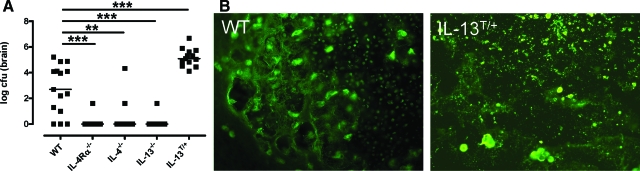
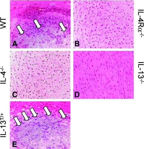
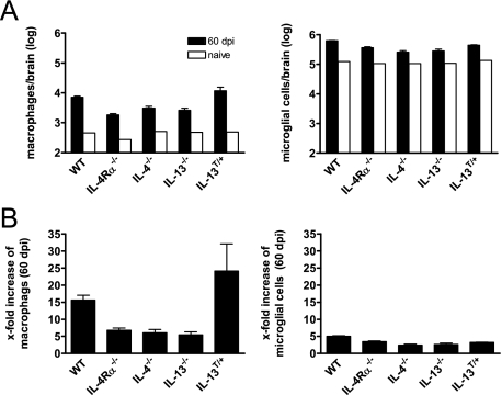
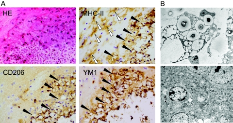
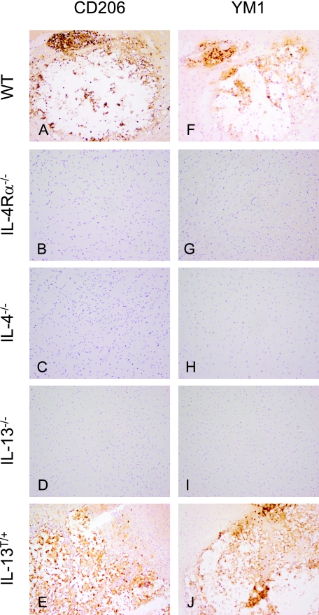
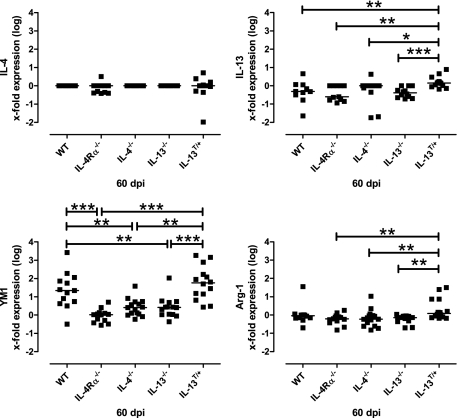
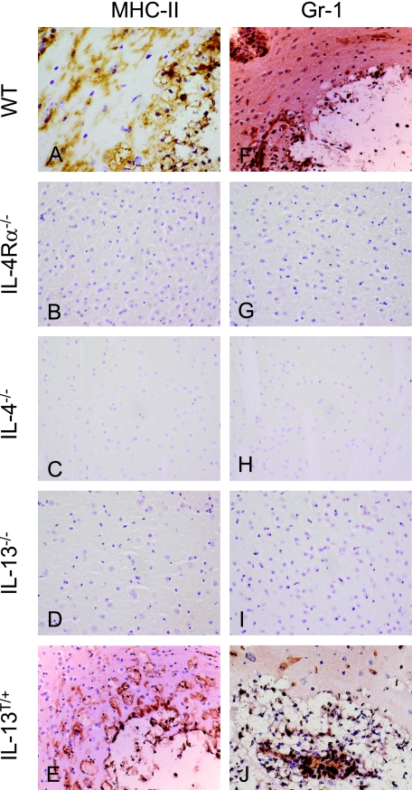
Both interleukin (IL)-4- and IL-13-dependent Th2-mediated immune mechanisms exacerbate murine Cryptococcus neoformans-induced bronchopulmonary disease. To study the roles of IL-4 and IL-13 in cerebral cryptococcosis, IL-4 receptor alpha-deficient (IL-4Ralpha(-/-)), IL-4-deficient (IL-4(-/-)), IL-13-deficient (IL-13(-/-)), IL-13 transgenic (IL-13(T/+)), and wild-type mice were infected intranasally. IL-13(T/+) mice displayed a higher fungal brain burden than wild-type mice, whereas the brain burdens of IL-4Ralpha(-/-), IL-4(-/-), and IL-13(-/-) mice were significantly lower as compared with wild-type mice. On infection, 68% of wild-type mice and 88% of IL-13-overexpressing IL-13(T/+) mice developed significant cerebral lesions. In contrast, only a few IL-4Ralpha(-/-), IL-4(-/-), and IL-13(-/-) mice had small lesions in their brains. Furthermore, IL-13(T/+) mice harbored large pseudocystic lesions in the central nervous system parenchyma, bordered by voluminous foamy alternatively activated macrophages (aaMphs) that contained intracellular cryptococci, without significant microglial activation. In wild-type mice, aaMphs tightly bordered pseudocystic lesions as well, and these mice, in addition, showed microglial cell activation. Interestingly, in resistant IL-4(-/-), IL-13(-/-), and IL-4Ralpha(-/-) mice, no aaMphs were discernible. Microglial cells of all mouse genotypes neither internalized cryptococci nor expressed markers of alternative activation, although they displayed similar IL-4Ralpha expression levels as macrophages. These data provide the first evidence of the development of aaMphs in a central nervous system infectious disease model, pointing to distinct roles of macrophages versus microglial cells in the central nervous system immune response against C. neoformans.


Figures

Figure 1
Figure 1
IL-4/IL-13 expression leads to elevated intracerebral fungal load in mice infected i.n. with C. neoformans for 60 days. A: The intracerebral fungal load of wild-type (WT), IL-4Rα−/−, IL-4−/−, IL-13−/−, and IL-13T/+ mice was counted after sterile removal of the brain (n = 5 animals/genotype) and data from four experiments were pooled. Data represent the median of 15 animals per group. Statistical analysis between wild-type, IL-4Rα−/−, IL-4−/−, IL-13−/−, and IL-13T/+ mice was performed by use of the Mann-Whitney-U Test. **P < 0.01; ***P < 0.001. B: In wild-type mice, huge amounts of anti-GXM positive cryptococci and yeast fragments can be found within a pseudocystic lesion. In IL-13T/+ mice, a large pseudocystic lesion, harboring densely packed masses of small yeast fragments, and large grouped intact yeasts are shown. Day 60 p.i.; Anti-GXM immunostaining magnification = original ×400.
Figure 2
Figure 2
Development of pseudocystic lesion in large scale in the brains of susceptible C. neoformans-infected wild-type and IL-13T/+ mice, but the absence of IL-4/−13 is associated with occasional formation of microglial nodules, granulomas, or only small pseudocystic lesions. Shown are representative micrographs (see supplemental figures). A–E: Day 60 pi; H&E staining of the brain of wild-type (WT), IL-4Rα−/−, IL-4−/−, IL-13−/−, and IL-13T/+ mice; magnification = original ×400. In wild-type and IL-13T/+ mice large foamy macrophages bordering pseudocystic lesions filled with cryptococci (white arrows) could be found, whereas the brain parenchyma of IL-4Rα−/−, IL-4−/−, and IL-13−/− mice is unremarkable.
Figure 3
Figure 3
High numbers of macrophages infiltrate the CNS in the presence of IL-4/IL-13 following i.n. infection with C. neoformans. For fluorescence-activated cell sorting analysis of intracerebral leukocytes, brains of five mice per genotype were pooled following i.n. infection with C. neoformans in BALB/c wild-type (WT), IL-4Rα−/−, IL-4−/−, IL-13−/−, and IL-13T/+ mice (A). Three similar experiments from infected mice and one experiment from naïve mice are shown. Cells were isolated, stained for Gr-1, CD11b, CD45, and analyzed by flow cytometry. Data are expressed as numbers of cells of the respective cell type per brain. Numbers of the respective cell types of naïve mice are shown in open bars. In (B) the x-fold increase of macrophages and microglial cells during cryptococcosis (60 dpi) in comparison with naïve mice is shown.
Figure 4
Figure 4
In the presence of IL-4 and IL-13, macrophages but not microglial cells expressing markers of alternative activation are detected in the brains of susceptible BALB/c wild-type mice at 60 d.p.i. A: Wild-type (WT) mice show that large pseudocystic lesion, harboring many small yeasts and yeast fragments, is bordered by inflammatory leukocytes, consisting mainly of large rounded foamy macrophages (black arrowheads). H&E staining, magnification = original ×600. The large foamy rounded cells are identified as MHC class II+ macrophages tightly bordering the lesion. Activated MHC class II+ ramified microglial cells (white arrowheads) are demonstrated in the outer vicinity of the lesion. Anti-MHC class II immunostaining, magnification = original ×600. Large round cells strongly express the CD206 antibody, whereas the antibody against the mannose receptor did not detect ramified microglial cells. Anti-CD206 immunostaining; magnification = original ×600. Many round cells, but not microglial cells, bordering the lesion strongly express the YM1 antibody. Anti-YM1 immunostaining; magnification = original ×600. B: Upper panel: Ultrastructural analysis of the identified type of macrophages reveals that they harbor yeasts and yeast fragments within their cytoplasm, and that they also contain numerous small vacuoles corresponding to their ‘foamy’ appearance, which are phagolysosomes. Lower panel: The ultrastructural analysis of microglial cells identifies a cell with dense-staining nucleus and marginated chromatin free of cryptococci or vacuolated lysosomes. Electron microscopy; magnification = original ×3000.
Figure 5
Figure 5
Alternatively activated macrophages develop in the CNS of susceptible wild-type, and IL-13T/+ mice after i.n. infection with C. neoformans. A–E: Mannose receptor (CD206) expression as a marker for alternative activation of macrophages in the brain (60 dpi). In wild-type (WT) and IL-13T/+ mice, macrophages show a strong expression of CD206, whereas the receptor is absent on cells of IL-4Rα−/− (B), IL-4−/− (C), and IL-13−/− (D) mice. In no case were microglial cells CD206 positive. Anti-CD206 immunostaining, magnification = original ×400. F–J: Chitinase YM1 expression as a marker for alternative activation of myeloid leukocytes in the brain (60 dpi). In wild-type and IL-13T/+ mice, macrophages show a strong expression of YM1, whereas the molecule is absent in cells of IL-4Rα−/− (G), IL-4−/− (H), and IL-13−/− (I) mice. In no case microglial cells were YM1 positive. Anti-YM1 immunostaining; magnification = original ×400.
Figure 6
Figure 6
Differential transcription of IL-4, IL-13, YM1, and arginase-1 in wild-type, IL-4Rα−/−, IL-4−/−, IL-13−/−, and IL-13T/+ mice after i.n. infection with C. neoformans. YM1 mRNA expression in the brains of wild-type (WT) and IL-13T/+ mice (60 dpi) was significantly elevated as compared with non-susceptible Th2 mutant mice. Arginase-1 mRNA expression in the brains of IL-13T/+ mice (60 dpi) was significantly elevated as compared with non-susceptible Th2 mutant mice. No IL-4 expression was detected in the CNS of all genotypes. IL-13 expression was not detected in all genotypes except for IL-13T/+ mice. Surprisingly, we detected some mRNA in IL-13 mutant mice using the primers described above, which results from the use of the IL-13 KO targeting vector. Data from 8 to 12 representative mice per genotype are shown in the graph. Statistical analysis between wild-type, IL-4Rα−/−, IL-4−/−, IL-13−/−, and IL-13T/+ mice was performed by use of the Mann-Whitney-U Test, with: *P < 0.05; **P < 0.01, ***P < 0.001.
Figure 7
Figure 7
Innate cellular immune reaction pattern in wild-type, IL-4Rα−/−, IL-4−/−, IL-13−/−, and IL-13T/+ mice after i.n. infection with C. neoformans. A–E: In a BALB/c wild-type (WT) mouse (A), foamy rounded MHC class II+ macrophages harboring yeast fragments are tightly bordering the lesion. In an IL-13T/+ mice (E), voluminous macrophages harboring yeast fragments within their cytoplasm are demonstrated at the border of a pseudocystic lesion. In comparison with the wild-type (A), the lesion is less well demarcated by macrophages, which rather diffusely infiltrate the parenchyma. Microglial activation is a less prominent feature in an IL-13T/+ mice, as compared with the wild-type (A). MHC class II+ cells are generally absent in the CNS of IL-4R−/−, IL-4−/−, and IL-13−/− mice (B–D). Day 60 p.i.; Anti-MHC class II immunostaining; magnification = original ×400. F–J: In a BALB/c wild-type mouse (F), Gr-1+ granulocytes are detected at the border and in the center of the lesion. Granulocytes also dissociate the vessel wall, which is localized at the upper left. In an Il-13T/+ mice (J), many Gr-1+ granulocytes are stained in the center and at the border of the lesion. Gr-1+ cells are generally absent in the CNS of IL-4R−/−, IL-4−/−, and IL-13−/− mice (G–I). F–J: Day 60 p.i.; Anti-Gr-1 immunostaining; magnification = original ×400.

References

    1. Mitchell TG, Perfect JR. Cryptococcosis in the era of AIDS–100 years after the discovery of Cryptococcus neoformans. Clin Microbiol Rev. 1995;8:515–548. - PMC - PubMed
    1. Lortholary O, Nunez H, Brauner MW, Dromer F. Pulmonary cryptococcosis. Semin Respir Crit Care Med. 2004;25:145–157. - PubMed
    1. Malik R, Krockenberger MB, Cross G, Doneley R, Madill DN, Black D, McWhirter P, Rozenwax A, Rose K, Alley M, Forshaw D, Russell-Brown I, Johnstone AC, Martin P, O'Brien CR, Love DN. Avian cryptococcosis. Med Mycol. 2003;41:115–124. - PubMed
    1. McAdams HP, Rosado-de-Christenson ML, Lesar M, Templeton PA, Moran CA. Thoracic mycoses from endemic fungi: radiologic-pathologic correlation. Radiographics. 1995;15:255–270. - PubMed
    1. McAdams HP, Rosado-de-Christenson ML, Templeton PA, Lesar M, Moran CA. Thoracic mycoses from opportunistic fungi: radiologic-pathologic correlation. Radiographics. 1995;15:271–286. - PubMed

Publication types

MeSH terms

LinkOut - more resources