. 2009 Feb;174(2):586-94.
doi: 10.2353/ajpath.2009.080661. Epub 2009 Jan 15.

The low-density lipoprotein receptor-related protein 1 mediates tissue-type plasminogen activator-induced microglial activation in the ischemic brain

Affiliations

The low-density lipoprotein receptor-related protein 1 mediates tissue-type plasminogen activator-induced microglial activation in the ischemic brain

Chen Zhang et al. Am J Pathol. 2009 Feb.

Abstract

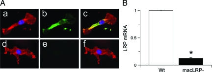
Microglia are the immune cells of the central nervous system (CNS) that become activated in response to pathological situations such as cerebral ischemia. Tissue-type plasminogen activator (tPA) is a serine proteinase that is found in the intravascular space and the CNS. The low-density lipoprotein receptor-related protein 1 (LRP1) is a member of the low-density lipoprotein receptor gene family found in neurons, astrocytes, and microglia. The present study investigated whether the interaction between tPA and microglial LRP1 plays a role in cerebral ischemia-induced microglial activation. We found that middle cerebral artery occlusion (MCAO) induces microglial activation in both wild-type and plasminogen-deficient (Plg(-/-)) mice. In contrast, MCAO-induced microglial activation is significantly decreased in tPA-deficient (tPA(-/-)) mice and in mice that lack LRP1 in microglial cells (macLRP(-)). We observed a significant increase in microglial activation when tPA(-/-) mice received treatment with murine tPA after MCAO. In contrast, treatment of macLRP(-) mice with tPA did not have an effect on the extent of microglial activation. Finally, both the volume of the ischemic lesion as well as inducible nitric oxide synthase production were significantly decreased in macLRP(-) mice and macLRP(-) microglia. In summary, our results indicate that the interaction between tPA and LRP1 induces microglial activation with the generation of an inflammatory response in the ischemic brain, suggesting a cytokine-like role for tPA in the CNS.


Figures

Figure 1
Figure 1
TPA mediates cerebral ischemia-induced microglial activation via a plasminogen-independent mechanism. A: Representative micrographs corresponding to immunohistochemical analysis of microglial activation in the AOI-2 24 hours after transient middle cerebral artery occlusion (tMCAO) in wild-type (a–d), and tPA−/− (e–h) mice, as well as in tPA−/− mice treated with tPA (i–l), and in Plg−/− mice (m–p). c, g, k, and o represent the merged images corresponding to the ipsilateral, ischemic hemisphere, whereas d, h, l, and p are the merged images for the corresponding area in the contralateral, nonischemic hemisphere. Blue is DAPI, red is F4/80, and green is β-isolectin. The insets in a–c correspond to a higher magnification of examples of cells with ameboid morphology and positive for β-isolectin and F4/80. Images were visualized using a Leica DMRBE microscope (Leica, Houston, TX) equipped with a 100×/1.30 numerical aperture (NA) and a LeicaDC500 camera. B: Average percentage of active microglial cells in each AOI 24 hours after tMCAO in wild-type (Wt, white bars), tPA−/− (black bars), and Plg−/− (gray bar) mice. atPA denotes treatment with active tPA whereas itPA denotes treatment with inactive tPA immediately after tMCAO. NT, no treatment. n = 10. Bars indicate SEM. NS, nonsignificant. Original magnifications: ×20 (a–p); ×40 (insets in a–c).
Figure 2
Figure 2
Microglial LRP1 is effectively deleted in macLRP mice. A: Representative micrograph of confocal microscopy analysis of microglial cultures from wild-type (a–c) and macLRP mice (d–f). Blue is DAPI, red is Mac-1, and green is LRP. c and f correspond to merged images. B: Quantitative RT-PCR analysis in microglial cultures from wild-type (Wt, white bar) and macLRP (black bar) mice. n = 4. Bars denote SD. *P < 0.001. Original magnifications, ×100.
Figure 3
Figure 3
LRP1 mediates tPA-induced microglial activation after MCAO. A: Representative micrographs corresponding to immunohistochemical analysis for β-isolectin and F4/80 in the AOI-2 24 hours after transient middle cerebral artery in wild-type (a–c) and macLRP (d–i) mice. g–i correspond to macLRP mice treated with tPA immediately after MCAO. c, f, and i represent the merged images. Blue is DAPI, red is F4/80, and green is β-isolectin. Images were visualized using a Leica DMRBE microscope equipped with a 100×/1.30 numerical aperture (NA) and a Leica DC500 camera. B: Average percentage of activated microglial cells in each AOI 24 hours after tMCAO in Wt (white bars) and macLRP (black bars) mice, either left untreated (NT) or intracerebrally injected with tPA immediately after MCAO (+tPA). n = 10. NS, nonsignificant. Original magnifications, ×20.
Figure 4
Figure 4
Effect of microglial LRP1 on the volume of the ischemic lesion. A: Example of 2,3,5-triphenyltetrazolium chloride staining in wild-type (Wt) and macLRP mice 24 hours after MCAO. The black arrows depict the infracted area. B: Volume of the ischemic lesion 24 hours after tMCAO in Wt and macLRP mice. n = 10; *P < 0.001. Bars depict SEM.
Figure 5
Figure 5
Role of microglial LRP1 on cerebral ischemia-induced iNOS expression. A and B: Quantitative real-time RT-PCR analysis of iNOS expression in microglial cultures 3 hours after exposure to oxygen-glucose deprivation conditions (A) and brain extracts 24 hours after MCAO (B). NT, no treatment; +tPA, animals treated with tPA immediately after MCAO; Wt, wild-type mice; macLRP, mice deficient in microglial LRP1. Error bars described SEM. n = 6. *P < 0.05 when compared with either macLRP microglia (A) or with untreated (NT) tPA mice or untreated (NT) and treated (+tPA) macLRP mice (B).
Figure 6
Figure 6
Role of microglial LRP1 on cerebral ischemia-induced nitrotyrosine accumulation. A: Representative pictures of immunohistochemical analysis for nitrotyrosine formation in the AOI-2 in wild-type (a–c) and macLRP (d–i) mice. g–i correspond to macLRP mice treated with tPA immediately after MCAO. c, f, and i are merged images. Blue is DAPI, red is F4/80, and green is nitrotyrosine. B: Western blot analysis of nitrotyrosine formation 24 hours after MCAO in wild-type (Wt) and macLRP mice either left untreated (NT) or intracerebrally injected with tPA (+tPA) immediately after MCAO. Original magnifications, ×20.

References

    1. del Rio-Hortega P. Microglia. Penfield W, editor. New York: Hoeber,; Cytology and Cellular Pathology of the Nervous System. 1932:pp 482–534.
    1. Hanisch UK, Kettenmann H. Microglia: active sensor and versatile effector cells in the normal and pathologic brain. Nat Neurosci. 2007;10:1387–1394. - PubMed
    1. Ladeby R, Wirenfeldt M, Garcia-Ovejero D, Fenger C, Dissing-Olesen L, Dalmau I, Finsen B. Microglial cell population dynamics in the injured adult central nervous system. Brain Res Rev. 2005;48:196–206. - PubMed
    1. Iadecola C, Zhang F, Casey R, Nagayama M, Ross ME. Delayed reduction of ischemic brain injury and neurological deficits in mice lacking the inducible nitric oxide synthase gene. J Neurosci. 1997;17:9157–9164. - PMC - PubMed
    1. Parathath SR, Parathath S, Tsirka SE. Nitric oxide mediates neurodegeneration and breakdown of the blood-brain barrier in tPA-dependent excitotoxic injury in mice. J Cell Sci. 2006;119:339–349. - PubMed

Publication types

MeSH terms