. 2009 Feb;5(2):82-90.
doi: 10.1038/nchembio.139. Epub 2009 Jan 15.

DNA damage tolerance: when it's OK to make mistakes

Affiliations

DNA damage tolerance: when it's OK to make mistakes

Debbie J Chang et al. Nat Chem Biol. 2009 Feb.

Abstract

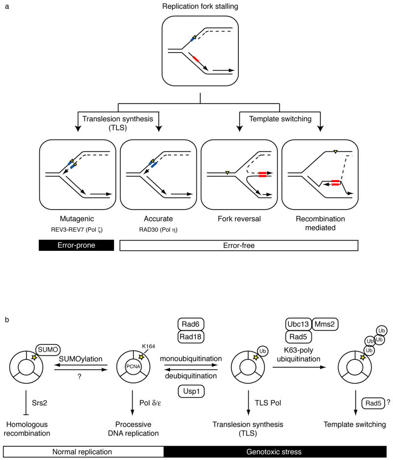
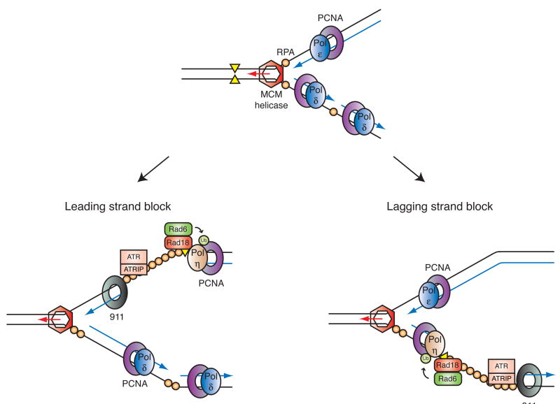
Mutations can be beneficial under conditions in which genetic diversity is advantageous, such as somatic hypermutation and antibody generation, but they can also be lethal when they disrupt basic cellular processes or cause uncontrolled proliferation and cancer. Mutations arise from inaccurate processing of lesions generated by endogenous and exogenous DNA damaging agents, and the genome is particularly vulnerable to such damage during S phase. In this phase of the cell cycle, many lesions in the DNA template block replication. Such lesions must be bypassed in order to preserve fork stability and to ensure completion of DNA replication. Lesion bypass is carried out by a set of error-prone and error-free processes collectively referred to as DNA damage tolerance mechanisms. Here, we discuss how two types of DNA damage tolerance, translesion synthesis and template switching, are regulated at stalled replication forks by ubiquitination of PCNA, and the conditions under which they occur.


Figures

Figure 1
Figure 1. Overview of DNA damage tolerance pathways and PCNA ubiquitination
(a) Lesions in the DNA template (yellow triangle) block processive DNA replication (dashed line). DNA damage tolerance mechanisms allow bypass of replication-blocking lesions by replicating over the damaged DNA (translesion synthesis, left) or using the undamaged sister chromatid (template switching, right). Template switching involves a structural rearrangement of the replication fork for which two models have been proposed. Fork reversal involves the formation of a four-way junction or “chicken-foot” intermediate (left) while recombination-mediated template switching involves D-loop formation and strand invasion (right). Templates used to bypass lesions and their complimentary sequences are boxed in blue for translesion synthesis and red for template switching. The mutagenic nature of the process is indicated. (b) Overview of functions for post-translationally modified forms of PCNA and the enzymes that carry out the modifications. In the absence of DNA damage, S. cerevisiae PCNA is SUMOylated at a conserved site (K164, yellow star). This modification allows recruitment of the helicase Srs2, which inhibits homologous recombination during normal replication. Although SUMOylation is a reversible process, deSUMOylation of PCNA has not yet been characterized. Following genotoxic stress, PCNA is ubiquitinated at K164 to promote DNA damage tolerance pathways. Monoubiquitinated PCNA facilitates translesion synthesis (TLS) through recruitment of TLS polymerases, while K63-linked (nondegradable) polyubiquitinated PCNA is associated with template switching, possibly utilizing the helicase activity of Rad5. Each ubiquitination step is mediated by a distinct set of enzymes. Rad6 and Ubc13-Mms2 are E2 ubiquitin-conjugating enzymes (green). Rad18 and Rad5 are E3 ubiquitin ligases (red). Usp1 is a deubiquitinating enzyme.
Figure 2
Figure 2. Translesion synthesis pathway
(a) Structures of a high fidelity replicative DNA polymerase and a low fidelity TLS polymerase (reprinted with permission from Nature Reviews Molecular & Cell Biology). The more restrictive nature of the active site in the replicative polymerase is apparent. (b) Model for polymerase switching during TLS. Replicative DNA polymerases (blue) stall at lesions (yellow triangle) in the DNA template. In the first polymerase switch, a specialized TLS polymerase (tan) is recruited to the sliding clamp PCNA at the stalled fork and replicates over the lesion. This TLS “patch” is then extended by the same or another TLS polymerase (green). The final switch restores the replicative DNA polymerase to the template and processive DNA synthesis continues. (c) Molecular interactions important for the switch from replicative to TLS polymerases. Following replication arrest, Rad6-Rad18 (E2–E3) ubiquitin enzymes are recruited to RPA bound single-stranded DNA. Following ubiquitination of PCNA by this complex, the TLS polymerase is recruited to the stalled fork. Polη interacts with three proteins at these sites via three different motifs: a ubiquitin binding domain (UBZ, green) which binds to the ubiquitin moiety on PCNA, a PIP box (purple) which binds to the hydrophobic pocket between the subunits of PCNA, and a carboxy-terminal domain (red) that mediates an interaction with Rad18. Whether the replicative polymerase (Polε) is actually displaced from PCNA as shown or moves aside and remains bound to PCNA is not clear.
Figure 3
Figure 3. Ubiquitin conjugation pathway and enzymes involved in PCNA ubiquitination
Cartoon depiction of the ubiquitin conjugation pathway. Ubiquitination occurs through at least three concerted reactions. Enzymes responsible for mono- and polyubiquitination of PCNA are as indicated. Human homologs of yeast Rad5 are in parenthesis. E1 = ubiquitin activating enzyme (yellow). E2 = ubiquitin conjugating enzyme (green). E3 = ubiquitin ligase (red). DUB = deubiquitinating enzyme (orange). Ub = ubiquitin. Substrate is shown in purple.
Figure 4
Figure 4. Domain architecture and functions of the E3 ubiquitin ligase Rad5 and its putative orthologs SHPRH and HLTF
(a) Structural comparisons between S. cerevisiae Rad5 and its putative human orthologs, HLTF and SHPRH. Blue and purple modules represent the seven helicase motifs characteristic of the SWI/SNF2 family of ATP-driven motor proteins. Although these motifs are spread over the length of the protein, they are collectively referred to as a helicase domain. Other domain names and descriptions are as listed through the NCBI conserved domain database and are described in the text. (b) The possible effects of helicase activity on a model homologous fork substrate. Unwinding and annealing of the nascent and parental DNA strands (bottom) results in fork regression and double-stranded DNA products, while unwinding alone (top) leads to single-stranded DNA products. Helicases known to exhibit these activities are listed in blue. (c) A possible mechanism for template switching mediated by fork reversal. Fork regression requires (i) concerted unwinding and annealing of the newly synthesized DNA strands, (ii) extension of the strand formed by the stalled polymerase past the sequence where the lesion is found on the parental template, and (iii) unwinding of the newly formed duplex so that the nascent strands can reanneal to their original templates and restore the fork to its proper conformation. Model four-way junction and homologous fork structures, which are known substrates of Rad5 in vitro, are placed in brackets adjacent to the fork structures they are thought to mimic. Yellow triangle = replication-blocking lesion. Dashed line = leading strand. Red box = template.
Figure 5
Figure 5. Coordinated activation of PCNA ubiquitination and the ATR-dependent checkpoint response at a stalled replication fork
Primed ssDNA accumulates at stalled replication forks when a polymerase stalls. In apparently independent processes, the resulting structure supports assembly of the proteins required for PCNA ubiquitination (Rad6 and Rad18) as well as the proteins required for activation of the ATR-dependent checkpoint pathway. For simplicity, only a few checkpoint proteins are depicted here: the 911 checkpoint clamp and the ATR-ATRIP heterodimer. Proper assembly of these proteins leads to phosphorylation of the downstream effector kinase Chk1 and cell cycle arrest. Shown are the presumed effects of stalling the leading (left) and lagging (right) strand polymerases. On the leading strand, ssDNA accumulates upon functional uncoupling of MCM helicase and replicative DNA polymerase activities, and in this case, the 5′-primer end thought to be required for checkpoint activation could be provided by the adjacent origin, or possibly replication restart. Note how a single stalled fork (i.e. gap) can be detected simultaneously by the two PCNA and 911 clamps.

References

    1. Friedberg EC. Suffering in silence: the tolerance of DNA damage. Nat Rev Mol Cell Biol. 2005;6:943–953. - PubMed
    1. Cimprich KA, Cortez D. ATR: an essential regulator of genome integrity. Nat Rev Mol Cell Biol. 2008;9:616–627. - PMC - PubMed
    1. Nyberg KA, Michelson RJ, Putnam CW, Weinert TA. Toward maintaining the genome: DNA damage and replication checkpoints. Annu Rev Genet. 2002;36:617–656. - PubMed
    1. Paulsen RD, Cimprich KA. The ATR pathway: fine-tuning the fork. DNA Repair (Amst) 2007;6:953–966. - PubMed
    1. Andersen PL, Xu F, Xiao W. Eukaryotic DNA damage tolerance and translesion synthesis through covalent modifications of PCNA. Cell Res. 2008;18:162–173. - PubMed

Substances