. 2008:2008:298010.
doi: 10.1155/2008/298010. Epub 2009 Jan 14.

Suppressive effect of hydroquinone, a benzene metabolite, on in vitro inflammatory responses mediated by macrophages, monocytes, and lymphocytes

Affiliations

Suppressive effect of hydroquinone, a benzene metabolite, on in vitro inflammatory responses mediated by macrophages, monocytes, and lymphocytes

Jae Youl Cho. Mediators Inflamm. 2008.

Abstract

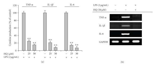
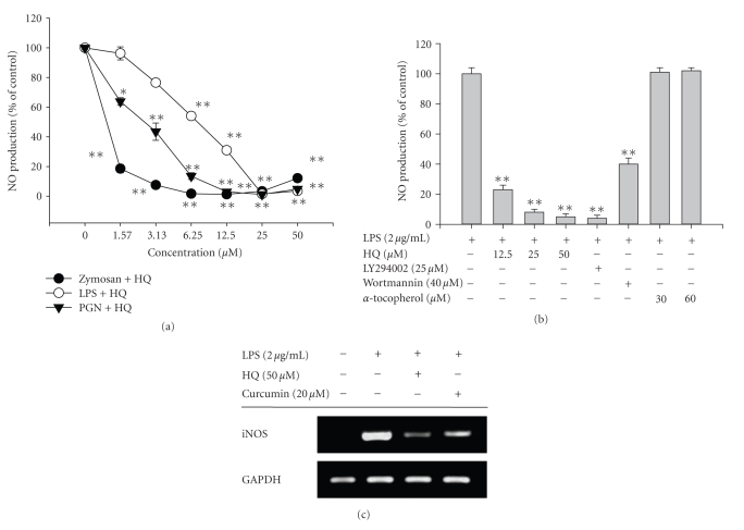
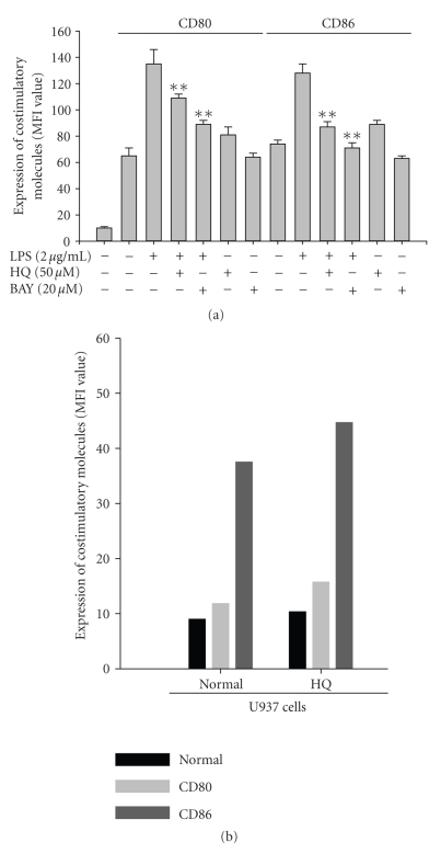
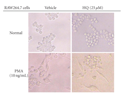
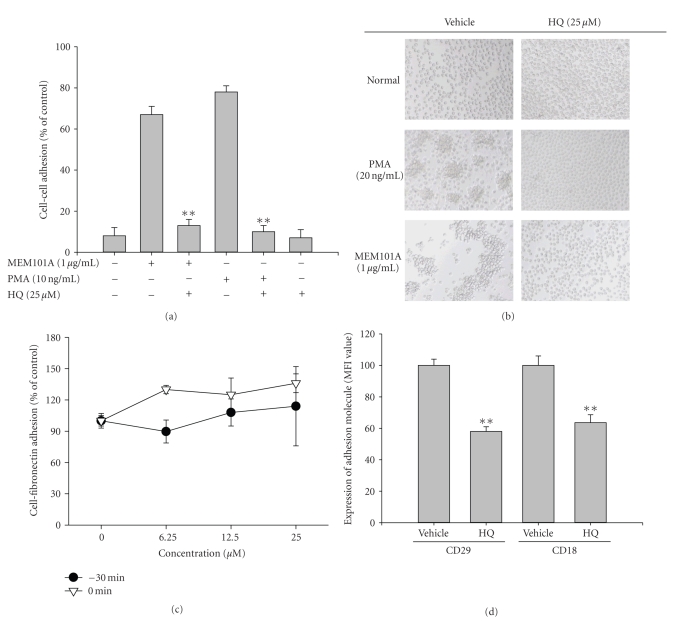
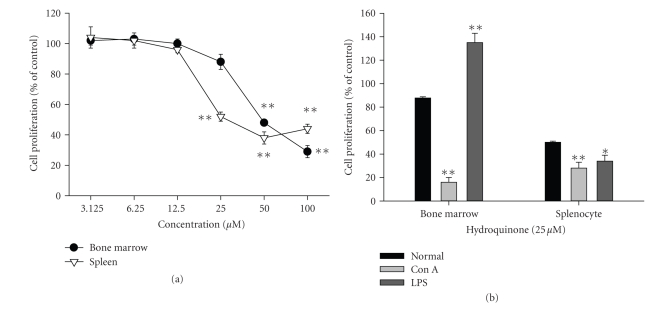
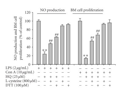
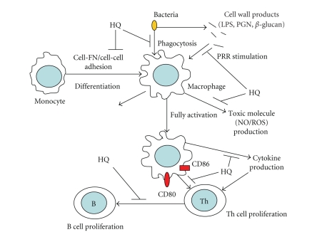
We investigated the inhibitory effects of hydroquinone on cytokine release, phagocytosis, NO production, ROS generation, cell-cell/cell fibronectin adhesion, and lymphocyte proliferation. We found that hydroquinone suppressed the production of proinflammatory cytokines [tumor necrosis factor (TNF)-alpha, interleukin (IL)-1beta, and IL-6], secretion of toxic molecules [nitric oxide (NO) and reactive oxygen species (ROS)], phagocytic uptake of FITC-labeled dextran, upregulation of costimulatory molecules, U937 cell-cell adhesion induced by CD18 and CD29, and the proliferation of lymphocytes from the bone marrow and spleen. Considering that (1) environmental chemical stressors reduce the immune response of chronic cigarette smokers and children against bacterial and viral infections and that (2) workers in petroleum factories are at higher risk for cancer, our data suggest that hydroquinone might pathologically inhibit inflammatory responses mediated by monocytes, macrophages, and lymphocytes.


Figures

Figure 1
Figure 1
Chemical structure of hydroquinone.
Figure 2
Figure 2
Effect of hydroquinone on the production of cytokines and their mRNA expression in LPS-activated RAW264.7 cells. (a) RAW264.7 cells (2 × 10⁶ cells/mL) were incubated with indicated concentrations of HQ (hydroquinone) in the presence or absence of LPS (2 μg/mL) for 12 hours. Secreted levels of TNF-α, IL-1β, and IL-6 in culture supernatant were determined by ELISA. (b) The mRNA levels of proinflammatory cytokines from the RAW264.7 cells were determined by semiquantitative RT-PCR. The results show one representative experiment of three. *p < .05 and **p < .01 represent significant difference as compared to control.
Figure 3
Figure 3
Effects of hydroquinone on the production of NO in LPS-treated RAW 264.7 cells or peritoneal macrophages. (a) and (b) RAW264.7 cells or peritoneal macrophages (1 × 10⁶) were pretreated with various concentrations of HQ (hydroquinone) in the presence or absence of LPS (2 μg/mL), PGN (10 μg/mL), or zymosan (400 μg/mL) for 24 hours. The level of NO was determined by Griess reagent as described in Section 2. (c) The mRNA levels of proinflammatory cytokines from RAW264.7 cells were determined by semiquantitative RT-PCR. The results show one representative experiment of three. *p < .05 and **p < .01 represent significant difference as compared to control.
Figure 4
Figure 4
Effects of hydroquinone on ROS generation in SNP-induced RAW 264.7 cells. (a) Antioxidant effect of hydroquinone was examined by DPPH assay as described in Section 2. (b) RAW264.7 cells (1 × 10⁶) were pretreated with various concentrations of HQ (hydroquinone) in the presence or absence of SNP (100 μM) for 30 minutes. The level of generated ROS was determined by flowcytometric analysis as described in Section 2 . *p < .05 and **p < .01 represent significant difference as compared to control.
Figure 5
Figure 5
Effect of hydroquinone on the phagocytic uptake of FITC-labeled dextran. RAW264.7 cells (1 × 10⁶) were incubated with in the indicated concentrations of HQ (hydroquinone) in the presence or absence of 1 mg/mL of FITC-labeled dextran for 30 minutes. The uptake of dextran was detected by flow cytometric analysis as described in Section 2. **p < .01 represents significant difference as compared to control.
Figure 6
Figure 6
Effect of hydroquinone on surface expression of costimulatory molecules. (a) and (b) RAW264.7 or U937 cells (1 × 10⁶) were incubated with indicated concentrations of HQ (hydroquinone) in the presence or absence of LPS (2 μg/mL) for 24 hours. The surface expression of CD80 and CD86 was determined by flow cytometric analysis as described in Section 2. **p < .01 represents significant difference as compared to control.
Figure 7
Figure 7
Effect of hydroquinone on PMA-induced morphological changes. RAW264.7 cells (1 × 10⁵) were incubated with indicated concentrations of HQ (hydroquinone) in the presence or absence of PMA (10 ng/mL) for 72 hours. The images of the cells in culture were obtained using an inverted phase contrast microscope attached to a video camera.
Figure 8
Figure 8
Effect of hydroquinone on cell-cell or cell-fibronectin adhesion events and surface levels of integrins (CD18 and CD29). (a) and (b) U937 cells (1 × 10⁶) were incubated with indicated concentrations of HQ (hydroquinone) in the presence or absence of a proaggregation (activating) antibody against CD29 (MEM101A (1 μg/mL)) for 2 hours. The extent of cell-cell aggregation was determined by quantitative cell-cell adhesion assay as described in Materials and Methods. (b) Images of cells in culture were obtained using an inverted phase contrast microscope attached to a video camera. (c) U937 cells pretreated with hydroquinone were seeded on fibronectin (50 μg/mL)-coated plates and further incubated for 3 hours. Attached cells were determined by crystal violet assay, as described in Section 2. (d) U937 cells were incubated with hydroquinone (12.5 μM) for 3 hours. The surface expression of adhesion molecules (CD18 and CD29) was determined by flow cytometric analysis as described in Section 2. **p < .01 represents significant difference as compared to control.
Figure 9
Figure 9
Effect of hydroquinone on the viablility or proliferation of splenic and bone marrow-derived lymphocytes induced by Con A and LPS. (a) Splenic lymphocytes (5 × 10⁶ cells/mL) were incubated with hydroquinone under normal culture conditions. (b) Bone marrow-derived lymphocytes (5 × 10⁶ cells/mL) were incubated with hydroquinone in the presence or absence of Con A (1 μg/mL) and LPS (10 μg/mL). Cell viability or proliferation was determined by MTT assay. *p < .05 and **p < .01 represent significant difference as compared to normal or control.
Figure 10
Figure 10
Effect of thiol-containing compounds on hydroquione-mediated inhibition of NO production and lymphocyte proliferation. (a) RAW264.7 cells (1 × 10⁶) were pretreated with L-cysteine (800 μM) or DTT (100 μM) and HQ (hydroquinone, 25 μM) in the presence or absence of LPS (2.5 μg/mL) for 24 hours. The level of NO was determined by Griess reagent as described in Section 2. (b) Bone marrow-derived cells were incubated with L-cysteine (800 μM) or DTT (100 μM) and hydroquinone (25 μM) in the presence or absence of Con A (1 μg/mL) for 24 hours. **p < .01 and ## p < .01 represent significant difference as compared to control or HQ-treated group.
Figure 11
Figure 11
Schematic diagram of hydroquinone-mediated inhibition.

References

    1. Savelkoul HFJ, Neijens HJ. Immune responses during allergic sensitization and the development of atopy. Allergy. 2000;55(11):989–997. - PubMed
    1. Rana SVS, Verma Y. Biochemical toxicity of benzene. Journal of Environmental Biology. 2005;26(2):157–168. - PubMed
    1. Deisinger PJ, Hill TS, English JC. Human exposure to naturally occurring hydroquinone. Journal of Toxicology and Environmental Health. 1996;47(1):31–46. - PubMed
    1. Moerloose KB, Pauwels RA, Joos GF. Short-term cigarette smoke exposure enhances allergic airway inflammation in mice. American Journal of Respiratory and Critical Care Medicine. 2005;172(2):168–172. - PubMed
    1. Kim E, Kang BY, Kim TS. Inhibition of interleukin-12 production in mouse macrophages by hydroquinone, a reactive metabolite of benzene, via suppression of nuclear factor-κB binding activity. Immunology Letters. 2005;99(1):24–29. - PubMed

MeSH terms

LinkOut - more resources