. 2009 May-Jun;45(5-6):252-63.
doi: 10.1007/s11626-008-9162-5. Epub 2009 Jan 16.

PBX1 is dispensable for neural commitment of RA-treated murine ES cells

Affiliations

PBX1 is dispensable for neural commitment of RA-treated murine ES cells

Anne S Jürgens et al. In Vitro Cell Dev Biol Anim. 2009 May-Jun.

Abstract

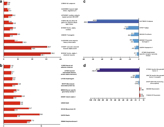
Experimentation with PBX1 knockout mice has shown that PBX1 is necessary for early embryogenesis. Despite broad insight into PBX1 function, little is known about the underlying target gene regulation. Utilizing the Cre-loxP system, we targeted a functionally important part of the homeodomain of PBX1 through homozygous deletion of exon-6 and flanking intronic regions leading to exon 7 skipping in embryonic stem (ES) cells. We induced in vitro differentiation of wild-type and PBX1 mutant ES cells by aggregation and retinoic acid (RA) treatment and compared their profiles of gene expression at the ninth day post-reattachment to adhesive media. Our results indicate that PBX1 interactions with HOX proteins and DNA are dispensable for RA-induced ability of ES to express neural genes and point to a possible involvement of PBX1 in the regulation of imprinted genes.


Figures

Figure 1.
Figure 1.
Schematic representation of the PBX1 locus modifications. (a) Schematic representation of the first allele targeting by homologous recombination and subsequent transient Cre recombinase expression. The targeting vector was based on the pFlox plasmid coding loxP flanked PGKNeo/HSV-TK selection cassette. The first targeting vector contained a 1191-nucleotide fragment of PBX1 locus bearing exon-6 flanked by loxP sequences. The HindIII site within the 5′ homology arm was removed from the targeting vector to allow homologous recombination detection by Southern blot. The 5′ external probe used for the Southern blot analysis is shown as a solid black box above the mutated allele. H HindIII, N NotI, Bg BglII, B BamHI, E EcoRI, S SalI. As a result of the first homologous recombination and subsequent transient Cre expression, the sequence containing exon-6 as well as the selection cassette was removed from the first allele, yielding neomycin-sensitive ΔPBX1+/− ES cell clones. (b) For the second targeting, the vector was modified by removing the target sequence containing exon-6 with BamHI. The second round of homologous recombination yielded neomycin-resistant ΔPBX1−/− clones.
Figure 2.
Figure 2.
Detection of targeted genetic modification of the PBX1 locus. (a) Southern blot analysis of PBX1 clones after the first homologous recombination, before Cre treatment. DNAs of individual clones resistant to G418 were digested with HindIII and analyzed by Southern hybridization using a PCR-derived 5′ external southern probe (for primer sequences, seeElectronic Supplementary Material). Shifts of WT 8-kb bands to 16-kb properly targeted allele bands were detected. (b) Confirmation of Southern blot analysis by long-range −8-kb PCR and standard −2-kb PCR on the 5′ and 3′ homology arms of the vector. In both PCRs, one primer was based on the sequences external to the targeting vector and one inside the vector sequence. (c) Southern blot analysis of the clones after Cre recombination using a 5′ external hybridization probe. Cre recombinase catalyzed removal of the selection cassette together with the target sequence resulted in a decrease of the original 16-kb band of the correctly targeted first allele to 11 kb ΔPBX1+/− band (Fig. 1c). The second homologous recombination resulted in a shift of the 8-kb WT allele band to the 14.5-kb correctly targeted second allele band.
Figure 3.
Figure 3.
Morphology of the wild-type E14, mutant ΔPBX1+/−, and ΔPBX1−/− ES cells growing undifferentiated on the embryonic fibroblast and differentiating upon aggregation in the presence of RA. (a, b) Low-magnification digital images of the colonies of undifferentiated WT and ΔPBX1−/− embryonic stem cells. (c, d) The same colonies photographed at higher magnification. WT and mutant cells grow in sharp-bordered colonies characteristic of undifferentiated ES cells. (e, f) Embryoid bodies formed by each of the ES cell clones as a result of aggregation in non-adhesive dishes followed by RA induction. (g, h) Morphology of differentiated ES cells 9 d after reattachment to the adhesive media. The ΔPBX1−/− cells have a flattened appearance as they spread in the surroundings of the reattached aggregates.
Figure 4.
Figure 4.
Analysis of PBX protein and mRNA expression. (a) Western blot analysis of PBX1/2/3 expression in the differentiated WT, ΔPBX1+/−, and ΔPBX1−/− cells on the ninth day after reattachment to adhesive cell culture dishes. Immunodetection of actin served as the loading control and was carried out on the same blots following PBX antibody striping. (b) Real-time PCR quantification of PBX2 and PBX3 transcripts in differentiated WT cells and two ΔPBX1−/− clones on the ninth day after reattachment to adhesive cell culture dishes.
Figure 5.
Figure 5.
The microarray-based comparative gene expression profiling of differentiated ΔPBX1−/− and WT cells. Represented are regulations consistently observed in two clones of ΔPBX1−/− cells. Upper and lower bars represent values of gene deregulation in ΔPBX1−/− clone-1 and ΔPBX1−/− clone-2, respectively, as compared to the WT cells. Fold change values are depicted next to the bars.
Figure 6.
Figure 6.
Differentiated ΔPBX1+/− and ΔPBX1−/− cells do not express IGF2, IGF2R, and colipase genes. Total RNA (15 μg/lane) isolated from undifferentiated (0) and differentiated (9) WT, ΔPBX1+/−, and ΔPBX1−/− embryonic stem cells was used in the Northern blot as described in “Materials and Methods”. Genotypes and stages of differentiation are assigned above the panel: (0) = undifferentiated, (9) = ninth day post-attachment of embryoid bodies to adhesive culture dishes. Efficiency of blotting was controlled by staining RNA on the membrane with ethidium bromide (top of the panel).
Figure 7.
Figure 7.
RA-treated ΔPBX1−/− ES cells differentiate into neurons. (a, b) Confocal microscopy images of differentiated WT and ΔPBX1−/− embryonic stem cells on the ninth day post-reattachment of embryoid bodies to adhesive media. (c, d) Expression of neuron-specific class III beta-tubulin (red) and extracellular signal-regulated kinases 1 (ERK1, green) were visualized by indirect immunofluorescence. A beta-III-tubilin composite of the same images. (e, f) FACS-based quantification of the beta-III-tubulin-stained, neural cell populations derived in WT (e) and ΔPBX1−/− (f) cell cultures.

References

    1. {'text': '', 'ref_index': 1, 'ids': [{'type': 'PubMed', 'value': '12376109', 'is_inner': True, 'url': 'https://pubmed.ncbi.nlm.nih.gov/12376109/'}]}
    2. Adamson S. L.; Lu Y.; Whiteley K. J.; Holmyard D.; Hemberger M.; Pfarrer C.; Cross J. C. Interactions between trophoblast cells and the maternal and fetal circulation in the mouse placenta. Dev. Biol. 250: 358–373; 2002. - PubMed
    1. {'text': '', 'ref_index': 1, 'ids': [{'type': 'DOI', 'value': '10.1016/j.ydbio.2003.09.041', 'is_inner': False, 'url': 'https://doi.org/10.1016/j.ydbio.2003.09.041'}, {'type': 'PubMed', 'value': '14732407', 'is_inner': True, 'url': 'https://pubmed.ncbi.nlm.nih.gov/14732407/'}]}
    2. Ahn J. I.; Lee K. H.; Shin D. M.; Shim J. W.; Lee J. S.; Chang S. Y.; Lee Y. S.; Brownstein M. J.; Lee S. H. Comprehensive transcriptome analysis of differentiation of embryonic stem cells into midbrain and hindbrain neurons. Dev. Biol. 265: 491–501; 2004. doi:10.1016/j.ydbio.2003.09.041. - PubMed
    1. {'text': '', 'ref_index': 1, 'ids': [{'type': 'DOI', 'value': '10.1016/S0169-328X(03)00256-0', 'is_inner': False, 'url': 'https://doi.org/10.1016/s0169-328x(03)00256-0'}, {'type': 'PubMed', 'value': '12941464', 'is_inner': True, 'url': 'https://pubmed.ncbi.nlm.nih.gov/12941464/'}]}
    2. Aihara K.; Kuroda S.; Kanayama N.; Matsuyama S.; Tanizawa K.; Horie M. A neuron-specific EGF family protein, NELL2, promotes survival of neurons through mitogen-activated protein kinases. Brain Res. Mol. Brain Res. 116: 86–93; 2003. doi:10.1016/S0169-328X(03)00256-0. - PubMed
    1. {'text': '', 'ref_index': 1, 'ids': [{'type': 'DOI', 'value': '10.1006/dbio.1995.1085', 'is_inner': False, 'url': 'https://doi.org/10.1006/dbio.1995.1085'}, {'type': 'PubMed', 'value': '7729574', 'is_inner': True, 'url': 'https://pubmed.ncbi.nlm.nih.gov/7729574/'}]}
    2. Bain G.; Kitchens D.; Yao M.; Huettner J. E.; Gottlieb D. I. Embryonic stem cells express neuronal properties in vitro. Dev. Biol. 168: 342–357; 1995. doi:10.1006/dbio.1995.1085. - PubMed
    1. {'text': '', 'ref_index': 1, 'ids': [{'type': 'PubMed', 'value': '2598811', 'is_inner': True, 'url': 'https://pubmed.ncbi.nlm.nih.gov/2598811/'}]}
    2. Beddington R. S.; Robertson E. J. An assessment of the developmental potential of embryonic stem cells in the midgestation mouse embryo. Development 105: 733–737; 1989. - PubMed

Publication types

MeSH terms

LinkOut - more resources