

Interactions between endothelial cells and electrospun methacrylic terpolymer fibers for engineered vascular replacements
- PMID: 19148926
- PMCID: PMC3432312
- DOI: 10.1002/jbm.a.32276
Interactions between endothelial cells and electrospun methacrylic terpolymer fibers for engineered vascular replacements
Abstract
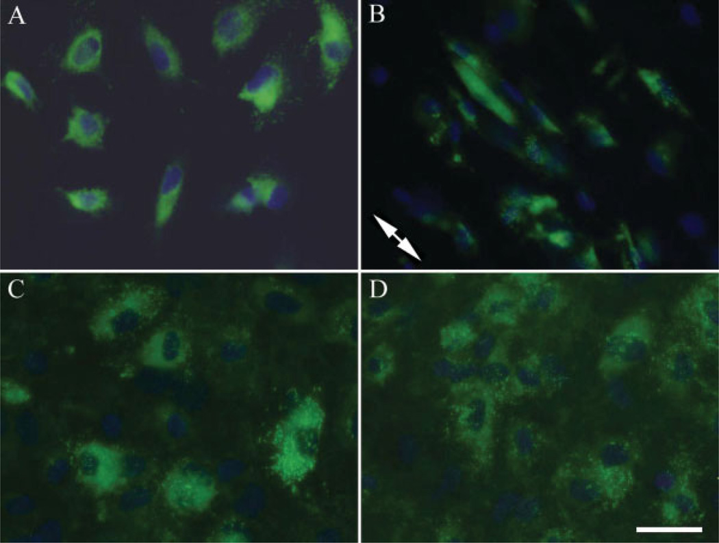
A compliant terpolymer made of hexylmethacrylate (HMA), methylmethacrylate (MMA), and methacrylic acid (MAA) intended for use in small diameter vascular graft applications has been developed. The mechanical properties and in vitro biostability of this terpolymer have been previously characterized. The goal of this investigation was to examine the interactions between endothelial cells and the new terpolymer and to evaluate endothelial cell function. Electrospinning was used to produce both oriented and random terpolymer fiber scaffolds. Smooth solution cast films and tissue culture polystyrene were used as negative and positive controls, respectively. Human blood outgrowth endothelial cells and human umbilical vein endothelial cells were incubated with the test and control samples and characterized with respect to initial cell attachment, proliferation, viability, and maintenance of the endothelial cell phenotype. It was found that the terpolymer is cytocompatible allowing endothelial cell growth, with random fibers being more effective in promoting enhanced cellular activities than oriented fibers. In addition, endothelial cells cultured on these substrates appeared to maintain their phenotype. The results from this study demonstrate that electrospun HMA:MMA:MAA terpolymer has the potential to be used successfully in fabricating small diameter blood vessel replacements.
Figures


References
-
- Lamba NMK, Woodhouse KA, Cooper SL. Polyurethanes in Biomedical Applications. Boca Raton, FL: CRC Press LLC; 1998.
-
- Boyd KL, Schmidt S, Pippert TR, Hite SA, Sharp WV. The effects of pore-size and endothelial-cell seeding upon the performance of small-diameter e-PTFE vascular grafts under controlled flow conditions. J Biomed Mater Res. 1988;22:163–177. - PubMed
-
- Vane JR, Anggard EE, Botting RM. Mechanisms of diseaseregulatory functions of the vascular endothelium. N Engl J Med. 1990;323:27–36. - PubMed
-
- Burkel WE, Graham LM, Stanley JC. Endothelial-linings in prosthetic vascular grafts. Ann N Y Acad Sci. 1987;516:131–144. - PubMed
-
- Lin HB, Garciaecheverria C, Asakura S, Sun W, Mosher DF, Cooper SL. Endothelial-cell adhesion on polyurethanes containing covalently attached RGD-peptides. Biomaterials. 1992;13:905–914. - PubMed
MeSH terms
Substances
Grants and funding
LinkOut - more resources
Full Text Sources
Other Literature Sources
Miscellaneous
