. 2009 Jan 16:8:4.
doi: 10.1186/1476-4598-8-4.

The chromatin remodelling factor BRG1 is a novel binding partner of the tumor suppressor p16INK4a

Affiliations

The chromatin remodelling factor BRG1 is a novel binding partner of the tumor suppressor p16INK4a

Therese M Becker et al. Mol Cancer. .

Abstract

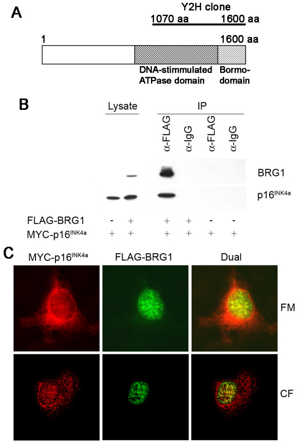
Background: CDKN2A/p16INK4a is frequently altered in human cancers and it is the most important melanoma susceptibility gene identified to date. p16INK4a inhibits pRb phosphorylation and induces cell cycle arrest, which is considered its main tumour suppressor function. Nevertheless, additional activities may contribute to the tumour suppressor role of p16INK4a and could help explain its specific association with melanoma predisposition. To identify such functions we conducted a yeast-two-hybrid screen for novel p16INK4a binding partners.

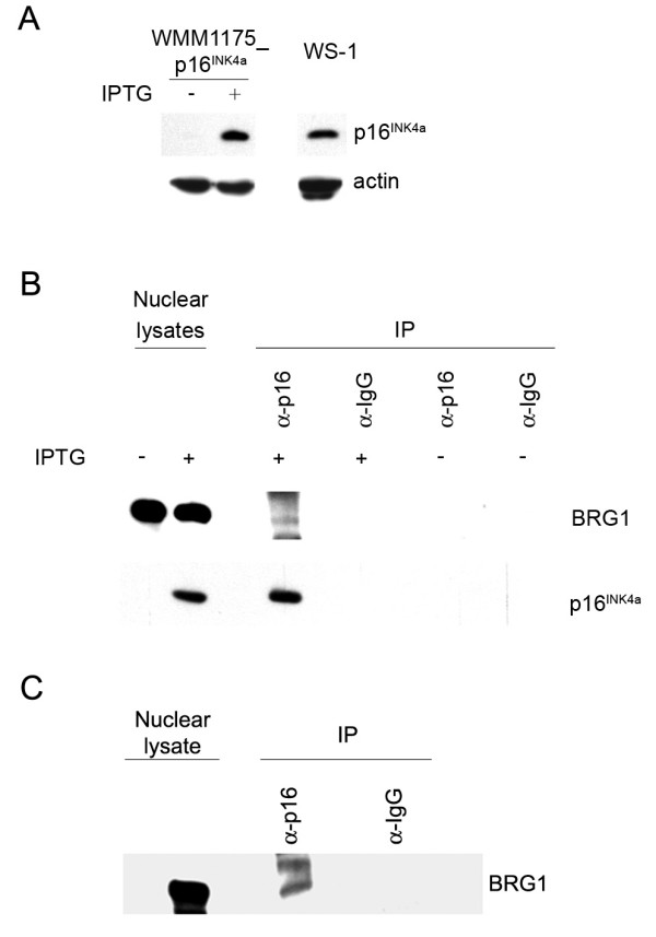
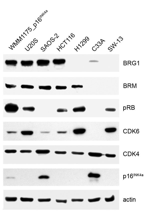
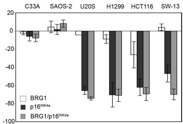
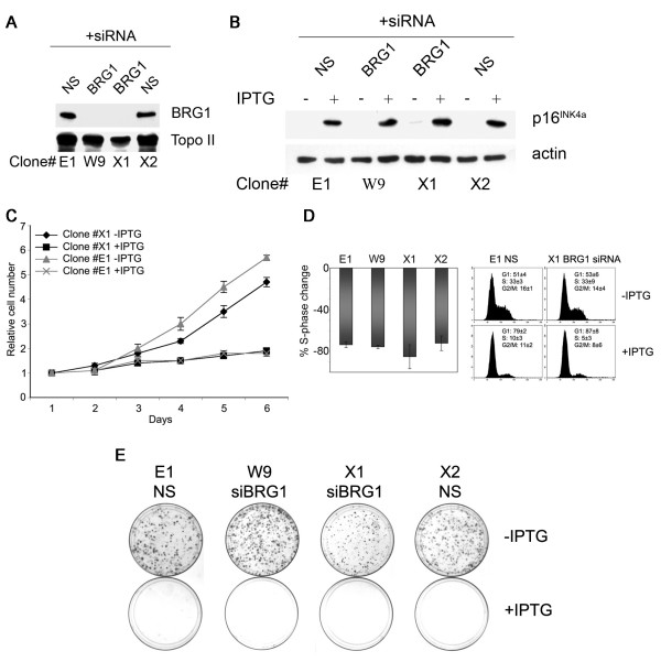
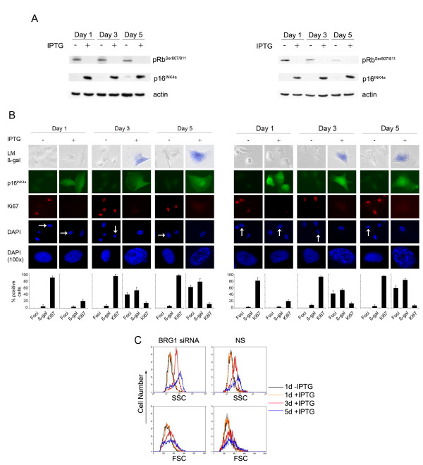
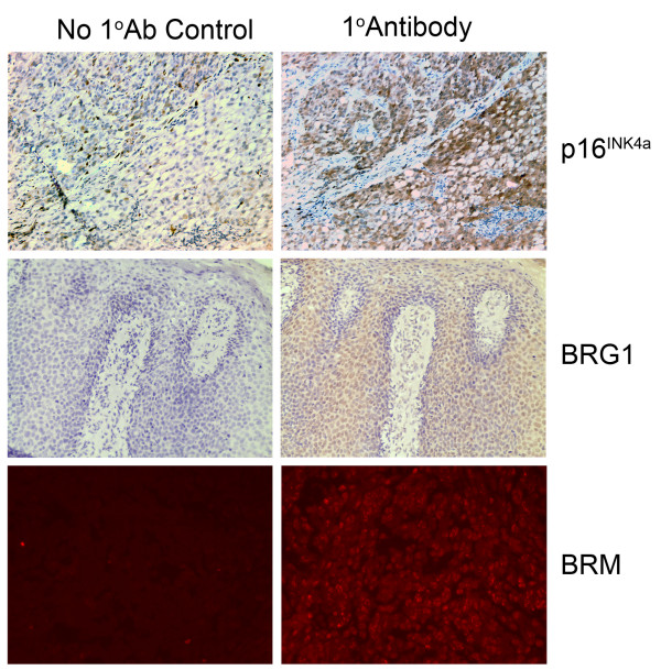
Results: We now report that p16INK4a interacts with the chromatin remodelling factor BRG1. We investigated the cooperative roles of p16INK4a and BRG1 using a panel of cell lines and a melanoma cell model with inducible p16INK4a expression and BRG1 silencing. We found evidence that BRG1 is not required for p16INK4a-induced cell cycle inhibition and propose that the p16INK4a-BRG1 complex regulates BRG1 chromatin remodelling activity. Importantly, we found frequent loss of BRG1 expression in primary and metastatic melanomas, implicating this novel p16INK4a binding partner as an important tumour suppressor in melanoma.

Conclusion: This data adds to the increasing evidence implicating the SWI/SNF chromatin remodelling complex in tumour development and the association of p16INK4a with chromatin remodelling highlights potentially new functions that may be important in melanoma predisposition and chemoresistance.


Figures

Figure 1
Figure 1
Identification of BRG1 as p16INK4a binding partner. A Schematic illustration of BRG1 highlighting the domains isolated in the yeast 2-hybrid screen (Y2H clone) B U2OS cells were transfected with MYC-p16INK4a and FLAG-BRG1 or control vector and immunoprecipitations were performed with a mouse-anti-FLAG antibody or a matched mouse IgG as indicated. BRG1 and p16INK4a were detected on immunoblots with anti-FLAG and anti-MYC antibodies. C Fluorescent microscopy images (FM) and confocal microscopy images (CF) of SW-13 cells grown on cover slips and transfected with MYC-p16INK4a and FLAG-BRG1 and probed with anti-FLAG and anti-MYC antibodies.
Figure 2
Figure 2
BRG1 binds p16INK4a in melanoma cells and normal fibroblasts. A 50 μg of total cell lysates derived from uninduced (-) and induced (+) WMM1175_p16INK4a cells and WS-1 fibroblasts (passage 20) were separated using a 15% SDS-PAGE gel. Immunoblots were probed for p16INK4a and β-actin as indicated. B WMM1175_p16INK4a cells were induced to express p16INK4a with 4 mM IPTG or mock treated for 72 hours. Immunoprecipitations were performed using a mouse anti-p16INK4a antibody or a matched mouse IgG from nuclear cell lysate, as indicated. Immunoblots were probed for endogenous BRG1 and induced p16INK4a using a mouse anti-BRG1 and rabbit anti-p16INK4a, respectively. C Endogenous BRG1 was co-immunoprecipitated with p16INK4a from WS-1 normal dermal human fibroblasts grown to passage 20 as detailed above.
Figure 3
Figure 3
pRb pathway proteins in cell lines. Expression of BRG1 and BRM was analyzed using 50 μg of nuclear cell lysates. All other proteins were analyzed from 50 μg of total cell lysates.
Figure 4
Figure 4
BRG1 and p16INK4a in cell cycle regulation. Indicated cell lines were transfected with MYC-p16INK4a, FLAG-BRG1 and/or a control vector plus GFP-spectrin. Cells were fixed with 70% ethanol 48 hours post transfection and cellular DNA was stained with propidium iodide. Percent S-phase change of GFP-spectrin positive cells was calculated (percent S-phase vector control - percent S-phase sample) × 100/percent S-phase vector control.
Figure 5
Figure 5
BRG1 does not alter p16INK4a cell cycle regulation. A 50 μg of nuclear lysates from WMM1175_p16INK4a clones with stably integrated siRNA targeting BRG1 or a non-specific (NS) control siRNA were probed for BRG1 and topoisomerase II (Topo II) as a loading control. B 50 μg of total cell lysates extracted from WMM1175_p16INK4a cells stably expressing either a BRG1-specific siRNA or a non-specific (NS) siRNA molecule, as indicated, were treated with PBS (-) or IPTG (+) for 24 h and probed for p16INK4a and β-actin. C Cell proliferation was determined by MTS assay. D A proportion of the IPTG/mock treated cells were analyzed for changes in cell cycle distribution. Percent S-phase change was calculated (percent S-phase mock treated cells – percent S-phase IPTG treated cells) × 100/percent S-phase mock treated cells. E The same clones were seeded at low density (103 cells/7.5 cm plate) and p16INK4a expression was induced with 4 mM IPTG or cells mock treated and colony forming ability was assayed after 14 days.
Figure 6
Figure 6
BRG1 does not alter p16INK4a driven senescence. WMM1175_p16INK4a cells, BRG1 silenced (clone X1, left panel) or NS (clone E1, right panel), were exposed to 4 mM IPTG over a five-day period and analyzed by FACS analysis, Western blot and imunocytostaining: A 50 μg of total cell lysate were immunoblotted and probed for p16INK4a, phospho-pRb (pRbSer807/811) and as a loading control β-actin. B The accumulation of p16INK4a, the cell proliferation marker Ki67, chromatin condensation (DAPI) and the appearance of SA-β-gal was analyzed by immunocytostaining in WMM1175_p16INK4a. Enlarged images of cells (indicated with arrows) show DAPI-stained chromatin foci. Histograms correspond to the average ± s.d of at least two independent induction experiments from a total of at least 500 cells. LM, light microscopy. C FACS analysis by Forward Scatter (FSC) and Side Scatter (SSC) of clones demonstrate the senescence associated increase of cell size (FSC) and granularity (SSC) upon p16INK4a induction.
Figure 7
Figure 7
Immunohistochemistry of melanomas for BRM, p16INK4a and BRG1. Melanoma samples were stained for p16INK4a and BRG1 with immunohistochemistry using DAB. BRM was stained using red fluorescence. Positive staining examples are presented in the right panel with no primary antibody control from the corresponding region in the left panel.

Similar articles

Cited by

References

    1. Goldstein AM, Chan M, Harland M, Gillanders EM, Hayward NK, Avril MF, Azizi E, Bianchi-Scarra G, Bishop DT, Bressac-de Paillerets B, et al. High-risk melanoma susceptibility genes and pancreatic cancer, neural system tumors, and uveal melanoma across GenoMEL. Cancer Res. 2006;66:9818–9828. doi: 10.1158/0008-5472.CAN-06-0494. - DOI - PubMed
    1. Sherr CJ, Roberts JM. CDK inhibitors: positive and negative regulators of G1-phase progression. Genes and Development. 1999;13:1501–1512. doi: 10.1101/gad.13.12.1501. - DOI - PubMed
    1. Stiegler P, De Luca A, Bagella L, Giordano A. The COOH-terminal region of pRb2/p130 binds to histone deacetylase 1 (HDAC1), enhancing transcriptional repression of the E2F-dependent cyclin A promoter. Cancer Research. 1998;58:5049–5052. - PubMed
    1. Muchardt C, Yaniv M. When the SWI/SNF complex remodels.the cell cycle. Oncogene. 2001;20:3067–3075. doi: 10.1038/sj.onc.1204331. - DOI - PubMed
    1. Zhang HS, Gavin M, Dahiya A, Postigo AA, Ma D, Luo RX, Harbour JW, Dean DC. Exit from G1 and S phase of the cell cycle is regulated by repressor complexes containing HDAC-Rb-hSWI/SNF and pRb-hSWI/SNF. Cell. 2000;101:79–89. doi: 10.1016/S0092-8674(00)80625-X. - DOI - PubMed

Publication types

MeSH terms

LinkOut - more resources