. 2009 Jul;12(6):805-22.
doi: 10.1017/S1461145708009802. Epub 2009 Jan 19.

Common effects of lithium and valproate on mitochondrial functions: protection against methamphetamine-induced mitochondrial damage

Affiliations

Common effects of lithium and valproate on mitochondrial functions: protection against methamphetamine-induced mitochondrial damage

Rosilla F Bachmann et al. Int J Neuropsychopharmacol. 2009 Jul.

Abstract

Accumulating evidence suggests that mitochondrial dysfunction plays a critical role in the progression of a variety of neurodegenerative and psychiatric disorders. Thus, enhancing mitochondrial function could potentially help ameliorate the impairments of neural plasticity and cellular resilience associated with a variety of neuropsychiatric disorders. A series of studies was undertaken to investigate the effects of mood stabilizers on mitochondrial function, and against mitochondrially mediated neurotoxicity. We found that long-term treatment with lithium and valproate (VPA) enhanced cell respiration rate. Furthermore, chronic treatment with lithium or VPA enhanced mitochondrial function as determined by mitochondrial membrane potential, and mitochondrial oxidation in SH-SY5Y cells. In-vivo studies showed that long-term treatment with lithium or VPA protected against methamphetamine (Meth)-induced toxicity at the mitochondrial level. Furthermore, these agents prevented the Meth-induced reduction of mitochondrial cytochrome c, the mitochondrial anti-apoptotic Bcl-2/Bax ratio, and mitochondrial cytochrome oxidase (COX) activity. Oligoarray analysis demonstrated that the gene expression of several proteins related to the apoptotic pathway and mitochondrial functions were altered by Meth, and these changes were attenuated by treatment with lithium or VPA. One of the genes, Bcl-2, is a common target for lithium and VPA. Knock-down of Bcl-2 with specific Bcl-2 siRNA reduced the lithium- and VPA-induced increases in mitochondrial oxidation. These findings illustrate that lithium and VPA enhance mitochondrial function and protect against mitochondrially mediated toxicity. These agents may have potential clinical utility in the treatment of other diseases associated with impaired mitochondrial function, such as neurodegenerative diseases and schizophrenia.


Figures
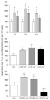
Fig. 1
Fig. 1
Effects of lithium (Li) and valproate (VPA) on cellular respiration in human SH-SY5Y cells. (a) Time-course of lithium and VPA effects on cellular respiration in SH-SY5Y cells. □, Control (Con); formula image, lithium; formula image, VPA. SH-SY5Y cells were exposed to lithium (1.2 mM) or VPA (1.0 mM) for 1, 3, or 6 d in serum-free, inositol-free Dulbecco’s Modified Eagle’s Medium. Cellular respiration was determined as described in the Methods section. (b) Dose–response curve for the effect of lithium on cellular respiration. (c) Dose–response curve for the effect of VPA on cellular respiration. Both lithium and VPA increased SH-SY5Y cell respiration rate at therapeutically relevant concentrations. Data are expressed as mean±S.E.M. (one-way ANOVA, N=3, n=5 for each group; Tukey’s multiple comparison test: * p<0.05).
Fig. 2
Fig. 2
Effects of lithium and valproate on mitochondrial membrane potential. SH-SY5Y cells were treated with various concentrations of lithium or valproate for 6 d and mitochondrial membrane potential was determined by JC-1 staining (one-way ANOVA, N=2, n=34; * p<0.05).
Fig. 3
Fig. 3
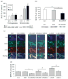
Lithium (Li) and valproate (VPA) enhance mitochondrial oxidation in SH-SY5Y cells. SH-SY5Y cells were treated with lithium (1.2 mM) or VPA (1.0 mM) for 3–7 d. The mitochondrial oxidation of VPA- or lithium-treated SH-SY5Y cells was determined by MitoTracker Red (MTR) staining. After 6–8 d of treatment, lithium (a, b) and VPA (c, d ) significantly enhanced mitochondrial oxidation (N=2–4, n=39–53, Student’s t test: * p<0.05). The MTR/MTG ratio showed similar changes (e). Data are expressed as mean±S.E.M.
Fig. 4
Fig. 4
Protective effect of lithium (Li) and valproate (VPA) on mitochondrial cytochrome c (Cyt c) and anti-apoptotic Bcl-2/Bax ratio in mitochondrial fractions after Meth treatment. Chronic treatment of rats with lithium or VPA attenuated Meth-induced decreases in anti-apoptotic Bcl-2 and increases in pro-apoptotic Bax in mitochondrial fraction of rat frontal cortex. Mitochondrial fraction was isolated by differential centrifugation (N=3, n=4–10, one-way ANOVA, Tukey’s multiple comparison test: ** p<0.01, * p<0.05; Student’s t test: # p<0.05). Data are expressed as mean±S.E.M. Porin was used as a loading control. (a) Representative blot and quantification of Bcl-2 changes in the mitochondrial fraction of rat frontal cortex. (b) Bax changes in the mitochondrial fraction of rat frontal cortex. (c) Ratio of Bcl-2/Bax in the mitochondrial fraction of rat frontal cortex. (d) Decrease of mitochondrial cytochrome c levels in rat frontal cortex after Meth administration, indicating that some of the cytochrome c was released into cytoplasm. Chronic treatment with lithium prevented Meth’s effects on cytochrome c levels. Quantified results are presented relative to controls.
Fig. 5
Fig. 5
Protective effects of lithium (Li) and valproate (VPA) on rat frontal cortex mitochondrial COX activity after Meth administration. Lithium and VPA preserved rat frontal cortex mitochondrial function by inhibiting the Meth-induced reduction of COX activity in frontal cortex homogenate. Cyclosporin A (CsA) was used as a positive control (N=2, n=6–28 animals per group; one-way ANOVA, Tukey’s multiple comparison test: * p<0.05; Student’s t test: # p<0.05).
Fig. 6
Fig. 6
Protective effect of lithium (Li) or valproate (VPA) on tyrosine hydroxylase (TH), a functional enzyme in dopaminergic neurons, after Meth injection in vivo. Chronic treatment of rats with lithium or VPA for 4 wk was followed by Meth injection for 1 d. Prefrontal cortical protein samples underwent Western blot analysis with anti-TH antibody. Actin was used as a loading control. Data are expressed as mean±S.E.M. (n=8 for each group, n=32; one-way ANOVA, Tukey’s multiple comparison test: ** p<0.05; Student’s t test: * p<0.05).
Fig. 7
Fig. 7
Protein expression levels (Western blot analysis) of five target genes selected from oligoarray analysis. Both lithium (Li) and valproate (VPA) pretreatment partially prevented Meth’s effects on the expression of five genes chosen for mitochondrial microarray profiling analysis. β-actin (ACTB) was used as a loading control. Mean ± S.E.M. are shown for relative protein levels based on densitometry analysis (n=8 animals for each group; one-way ANOVA and Tukey HSD post-hoc: * p<0.05, ** p<0.01. NDUFB10:NADH–ubiquinone oxidoreductase 1 beta subcomplex, 10).
Fig. 8
Fig. 8
Bcl-2 plays an important role in the effect of lithium (Li) and valproate (VPA) on mitochondrial function. (a) Lithium and VPA up-regulated Bcl-2 protein in SH-SY5Y cells in both total protein homogenates and mitochondrial fractions. SH-SY5Y cells of 80% confluency were treated with lithium (1.0 mM) or VPA (1.0 mM) for 6 d. Western blot analysis with anti-Bcl-2 antibody was performed to determine the protein level in cell homogenates and mitochondrial fraction. Data are expressed as mean ± S.E.M. (N=3, n=5 for each group; one-way ANOVA, Tukey’s multiple comparison test: * p<0.05). (b) Effects of Bcl-2 siRNA transfection on Bcl-2 protein levels. Bcl-2 siRNA significantly attenuated Bcl-2 expression in SH-SY5Y cells after 2 d of transfection. Data are expressed as mean ± S.E.M.; Student’s t test: * p<0.05 (n=4). (c) Effects of Bcl-2 knock-down with Bcl-2 siRNA on lithium- or VPA-evoked mitochondrial oxidation revealed by MitoTracker Red (MTR) staining. SH-SY5Y cells were exposed with or without VPA or lithium for 6–7 d after transfection with P-silencer containing siRNA sequence against Bcl-2 or scrambled sequence. MTR and MitoTracker Green (MTG) staining showed that Bcl-2 siRNA significantly attenuated mitochondrial oxidation in cultured SH-SY5Y cells; only the transfected cells were quantified as indicated by cyan fluorescent protein (CFP, blue). (d ) Bcl-2 siRNA, but not the scrambled siRNA (SCR), significantly attenuated MTR staining after treatment with VPA (1.0 mM) for 6 d or with lithium (1.2 mM) for 6 d (N=2–4, n=20–29; one-way ANOVA, Tukey’s multiple comparison test: # p<0.05; Student’s t test: * p<0.05). Data are expressed as mean ± S.E.M.

References

    1. Adams JM, Cory S. The Bcl-2 protein family: arbiters of cell survival. Science. 1998;281:1322–1326. - PubMed
    1. Adler CM, DelBello MP, Strakowski SM. Brain network dysfunction in bipolar disorder. CNS Spectrums. 2006;11:312–320. - PubMed
    1. Almeida A, Medina JM. A rapid method for the isolation of metabolically active mitochondria from rat neurons and astrocytes in primary culture. Brain Research. 1998;2:209–214. - PubMed
    1. Bar-Am O, Weinreb O, Amit T, Youdim MB. Regulation of Bcl-2 family proteins, neurotrophic factors, and APP processing in the neurorescue activity of propargylamine. FASEB Journal. 2005;19:1899–1901. - PubMed
    1. Belloc F, Belaud-Rotureau MA, Lavignolle V, Bascans E, Braz-Pereira E, Durrieu F, Lacombe F. Flow cytometry detection of caspase 3 activation in preapoptotic leukemic cells. Cytometry. 2000;40:151–160. - PubMed

Publication types

MeSH terms