

Phosphate-buffered saline-based nucleofection of primary endothelial cells
- PMID: 19150324
- PMCID: PMC2677097
- DOI: 10.1016/j.ab.2008.12.021
Phosphate-buffered saline-based nucleofection of primary endothelial cells
Abstract
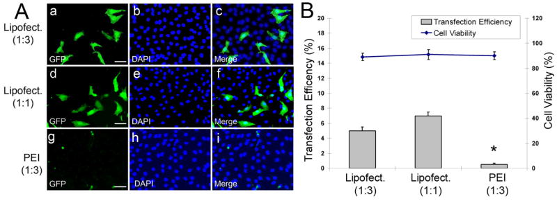
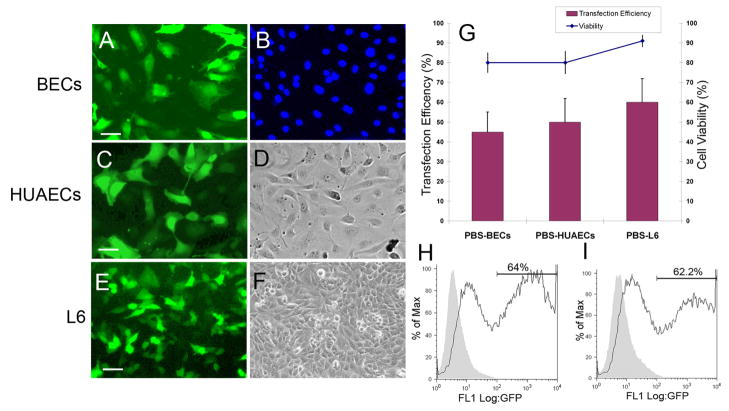
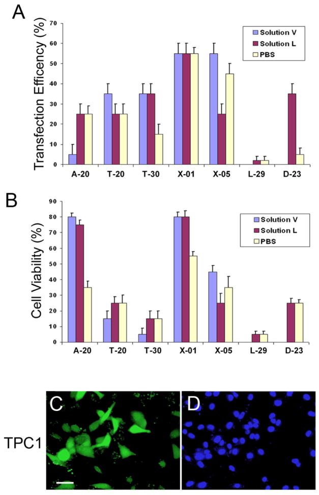
Although various nonviral transfection methods are available, cell toxicity, low transfection efficiency, and high cost remain hurdles for in vitro gene delivery in cultured primary endothelial cells. Recently, unprecedented transfection efficiency for primary endothelial cells has been achieved due to the newly developed nucleofection technology that uses a combination of novel electroporation condition and specific buffer components that stabilize the cells in the electrical field. Despite superior transfection efficiency and cell viability, high cost of the technology has discouraged cardiovascular researchers from liberally adopting this new technology. Here we report that a phosphate-buffered saline (PBS)-based nucleofection method can be used for efficient gene delivery into primary endothelial cells and other types of cells. Comparative analyses of transfection efficiency and cell viability for primary arterial, venous, microvascular, and lymphatic endothelial cells were performed using PBS. Compared with the commercial buffers, PBS can support equally remarkable nucleofection efficiency to both primary and nonprimary cells. Moreover, PBS-mediated nucleofection of small interfering RNA (siRNA) showed more than 90% knockdown of the expression of target genes in primary endothelial cells. We demonstrate that PBS can be an unprecedented economical alternative to the high-cost buffers or nucleofection of various primary and nonprimary cells.
Figures

Similar articles
-
The two hit hypothesis: an improved method for siRNA-mediated gene silencing in stimulated primary human T cells.J Immunol Methods. 2013 Oct 31;396(1-2):116-27. doi: 10.1016/j.jim.2013.08.005. Epub 2013 Aug 27. J Immunol Methods. 2013. PMID: 23988722
-
Nucleofection is a highly effective gene transfer technique for human melanoma cell lines.Exp Dermatol. 2008 May;17(5):405-11. doi: 10.1111/j.1600-0625.2007.00687.x. Epub 2008 Feb 27. Exp Dermatol. 2008. PMID: 18312380
-
Efficient gene delivery to primary alveolar epithelial cells by nucleofection.Am J Physiol Lung Cell Mol Physiol. 2013 Dec;305(11):L786-94. doi: 10.1152/ajplung.00191.2013. Epub 2013 Sep 27. Am J Physiol Lung Cell Mol Physiol. 2013. PMID: 24077946
-
Electroporation Knows No Boundaries: The Use of Electrostimulation for siRNA Delivery in Cells and Tissues.J Biomol Screen. 2015 Sep;20(8):932-42. doi: 10.1177/1087057115579638. Epub 2015 Apr 7. J Biomol Screen. 2015. PMID: 25851034 Free PMC article. Review.
-
siRNA delivery via electropulsation: a review of the basic processes.Methods Mol Biol. 2014;1121:81-98. doi: 10.1007/978-1-4614-9632-8_7. Methods Mol Biol. 2014. PMID: 24510814 Review.
Cited by
-
Phosphorylation of the transcription factor Sp4 is reduced by NMDA receptor signaling.J Neurochem. 2014 May;129(4):743-52. doi: 10.1111/jnc.12657. Epub 2014 Feb 12. J Neurochem. 2014. PMID: 24475768 Free PMC article.
-
Activated Transcription Factor 3 in Association with Histone Deacetylase 6 Negatively Regulates MicroRNA 199a2 Transcription by Chromatin Remodeling and Reduces Endothelin-1 Expression.Mol Cell Biol. 2016 Oct 28;36(22):2838-2854. doi: 10.1128/MCB.00345-16. Print 2016 Nov 15. Mol Cell Biol. 2016. PMID: 27573019 Free PMC article.
-
Development of a bio-electrospray system for cell and non-viral gene delivery.RSC Adv. 2018 Feb 9;8(12):6452-6459. doi: 10.1039/c7ra12477e. eCollection 2018 Feb 6. RSC Adv. 2018. PMID: 35540421 Free PMC article.
-
TGF-β regulates β-catenin signaling and osteoblast differentiation in human mesenchymal stem cells.J Cell Biochem. 2011 Jun;112(6):1651-60. doi: 10.1002/jcb.23079. J Cell Biochem. 2011. PMID: 21344492 Free PMC article.
-
Transcription factor SP4 phosphorylation is altered in the postmortem cerebellum of bipolar disorder and schizophrenia subjects.Eur Neuropsychopharmacol. 2015 Oct;25(10):1650-1660. doi: 10.1016/j.euroneuro.2015.05.006. Epub 2015 May 21. Eur Neuropsychopharmacol. 2015. PMID: 26049820 Free PMC article.
References
-
- Ear T, Giguere P, Fleury A, Stankova J, Payet MD, Dupuis G. High efficiency transient transfection of genes in human umbilical vein endothelial cells by electroporation. J Immunol Methods. 2001 Nov 1;257(1–2):41–9. - PubMed
-
- Kotnis RA, Thompson MM, Eady SL, Budd JS, Bell PR, James RF. Optimisation of gene transfer into vascular endothelial cells using electroporation. Eur J Vasc Endovasc Surg. 1995 Jan;9(1):71–9. - PubMed
-
- Nathwani AC, Gale KM, Pemberton KD, Crossman DC, Tuddenham EG, McVey JH. Efficient gene transfer into human umbilical vein endothelial cells allows functional analysis of the human tissue factor gene promoter. Br J Haematol. 1994 Sep;88(1):122–8. - PubMed
-
- Sipehia R, Martucci G. High-efficiency transformation of human endothelial cells by Apo E-mediated transfection with plasmid DNA. Biochem Biophys Res Commun. 1995 Sep 5;214(1):206–11. - PubMed
-
- Tanner FC, Carr DP, Nabel GJ, Nabel EG. Transfection of human endothelial cells. Cardiovasc Res. 1997 Sep;35(3):522–8. - PubMed
Publication types
MeSH terms
Substances
Grants and funding
LinkOut - more resources
Full Text Sources
