

Identification of the SSB binding site on E. coli RecQ reveals a conserved surface for binding SSB's C terminus
- PMID: 19150358
- PMCID: PMC2735845
- DOI: 10.1016/j.jmb.2008.12.065
Identification of the SSB binding site on E. coli RecQ reveals a conserved surface for binding SSB's C terminus
Abstract
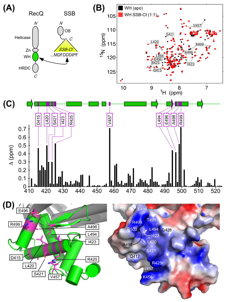
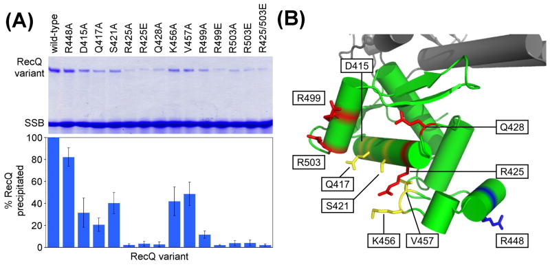
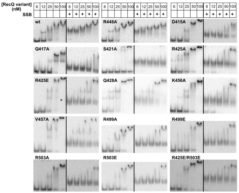
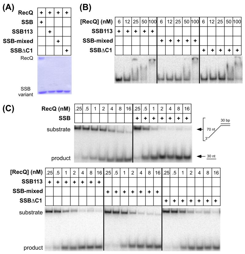
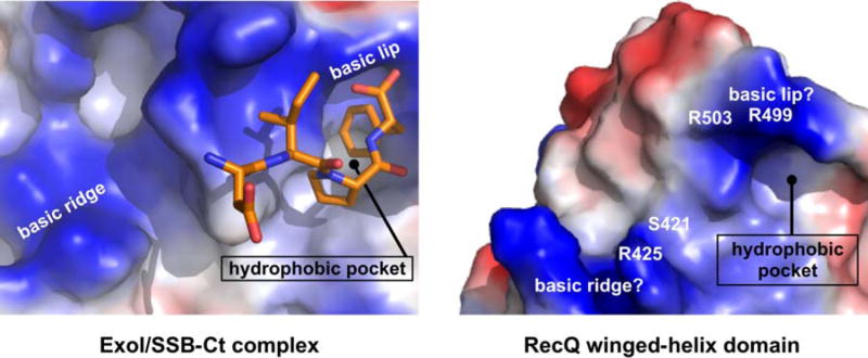
RecQ DNA helicases act in conjunction with heterologous partner proteins to catalyze DNA metabolic activities, including recombination initiation and stalled replication fork processing. For the prototypical Escherichia coli RecQ protein, direct interaction with single-stranded DNA-binding protein (SSB) stimulates its DNA unwinding activity. Complex formation between RecQ and SSB is mediated by the RecQ winged-helix domain, which binds the nine C-terminal-most residues of SSB, a highly conserved sequence known as the SSB-Ct element. Using nuclear magnetic resonance and mutational analyses, we identify the SSB-Ct binding pocket on E. coli RecQ. The binding site shares a striking electrostatic similarity with the previously identified SSB-Ct binding site on E. coli exonuclease I, although the SSB binding domains in the two proteins are not otherwise related structurally. Substitutions that alter RecQ residues implicated in SSB-Ct binding impair RecQ binding to SSB and SSB/DNA nucleoprotein complexes. These substitutions also diminish SSB-stimulated DNA helicase activity in the variants, although additional biochemical changes in the RecQ variants indicate a role for the winged-helix domain in helicase activity beyond SSB protein binding. Sequence changes in the SSB-Ct element are sufficient to abolish interaction with RecQ in the absence of DNA and to diminish RecQ binding and helicase activity on SSB/DNA substrates. These results support a model in which RecQ has evolved an SSB-Ct binding site on its winged-helix domain as an adaptation that aids its cellular functions on SSB/DNA nucleoprotein substrates.
Figures
Similar articles
-
A central role for SSB in Escherichia coli RecQ DNA helicase function.J Biol Chem. 2007 Jun 29;282(26):19247-58. doi: 10.1074/jbc.M608011200. Epub 2007 May 3. J Biol Chem. 2007. PMID: 17483090
-
RecQ helicase triggers a binding mode change in the SSB-DNA complex to efficiently initiate DNA unwinding.Nucleic Acids Res. 2017 Nov 16;45(20):11878-11890. doi: 10.1093/nar/gkx939. Nucleic Acids Res. 2017. PMID: 29059328 Free PMC article.
-
Single-strand DNA-binding protein suppresses illegitimate recombination in Escherichia coli, acting in synergy with RecQ helicase.Sci Rep. 2024 Sep 3;14(1):20476. doi: 10.1038/s41598-024-70817-5. Sci Rep. 2024. PMID: 39227621 Free PMC article.
-
SSB and the RecG DNA helicase: an intimate association to rescue a stalled replication fork.Protein Sci. 2017 Apr;26(4):638-649. doi: 10.1002/pro.3114. Epub 2017 Mar 17. Protein Sci. 2017. PMID: 28078722 Free PMC article. Review.
-
Molecular insights into the prototypical single-stranded DNA-binding protein from E. coli.Crit Rev Biochem Mol Biol. 2024 Feb-Apr;59(1-2):99-127. doi: 10.1080/10409238.2024.2330372. Epub 2024 May 21. Crit Rev Biochem Mol Biol. 2024. PMID: 38770626 Free PMC article. Review.
Cited by
-
Structure and Function of the PriC DNA Replication Restart Protein.J Biol Chem. 2016 Aug 26;291(35):18384-96. doi: 10.1074/jbc.M116.738781. Epub 2016 Jul 5. J Biol Chem. 2016. PMID: 27382050 Free PMC article.
-
The mechanism of Single strand binding protein-RecG binding: Implications for SSB interactome function.Protein Sci. 2020 May;29(5):1211-1227. doi: 10.1002/pro.3855. Epub 2020 Apr 17. Protein Sci. 2020. PMID: 32196797 Free PMC article.
-
Multiple C-terminal tails within a single E. coli SSB homotetramer coordinate DNA replication and repair.J Mol Biol. 2013 Nov 29;425(23):4802-19. doi: 10.1016/j.jmb.2013.08.021. Epub 2013 Sep 7. J Mol Biol. 2013. PMID: 24021816 Free PMC article.
-
Crystal structure of the Redβ C-terminal domain in complex with λ Exonuclease reveals an unexpected homology with λ Orf and an interaction with Escherichia coli single stranded DNA binding protein.Nucleic Acids Res. 2019 Feb 28;47(4):1950-1963. doi: 10.1093/nar/gky1309. Nucleic Acids Res. 2019. PMID: 30624736 Free PMC article.
-
RecQ helicase and RecJ nuclease provide complementary functions to resect DNA for homologous recombination.Proc Natl Acad Sci U S A. 2014 Dec 2;111(48):E5133-42. doi: 10.1073/pnas.1420009111. Epub 2014 Nov 19. Proc Natl Acad Sci U S A. 2014. PMID: 25411316 Free PMC article.
References
Publication types
MeSH terms
Substances
Grants and funding
LinkOut - more resources
Full Text Sources
Molecular Biology Databases
