. 2009 Apr 10;302(1):65-72.
doi: 10.1016/j.mce.2008.12.010. Epub 2008 Dec 25.

Cytoplasmic localization of the androgen receptor is independent of calreticulin

Affiliations

Cytoplasmic localization of the androgen receptor is independent of calreticulin

Minh M Nguyen et al. Mol Cell Endocrinol. .

Abstract

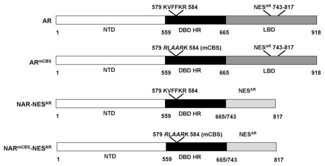
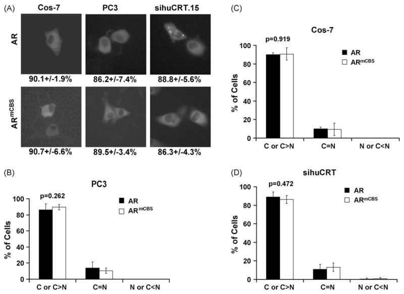
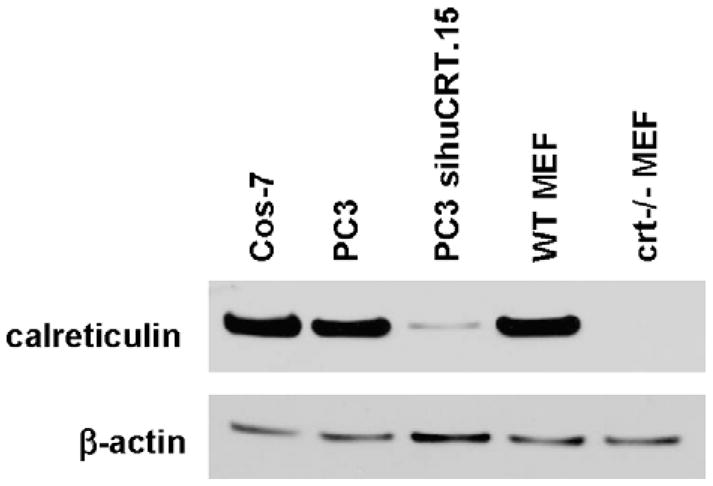
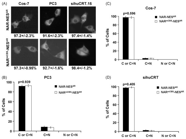
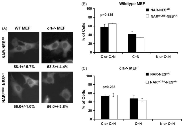
Identification and characterization of factors regulating intracellular localization of the androgen receptor (AR) are fundamentally important because nucleocytoplasmic trafficking of AR is a critical step in AR regulation by androgen manipulation. Normally, AR is localized to the cytoplasm in the absence of androgen. Upon ligand binding, AR translocates to the nucleus, where it can modulate transcription of AR-responsive genes. The withdrawal of androgen results in the export of unliganded AR from the nucleus to the cytoplasm, where it is transcriptionally inactive. Calreticulin has been implicated as a possible nuclear export factor for AR because the two proteins form a complex. In this study, we assessed whether the cytoplasmic localization of AR requires binding to calreticulin. To test this we substituted the calreticulin binding sequence (CBS) KVFFKR (residues 579-584) with the amino acids RLAARK in AR and monitored the cellular localization of a GFP-AR fusion protein in the absence of androgen. We also determined if knockdown or knockout of calreticulin expression affected the cytoplasmic localization of the AR. We found that a mutated CBS did not affect the localization of AR and that in the absence of androgen, AR is localized to the cytoplasm regardless of its ability to interact with calreticulin. Also, a reduction in the levels or loss of calreticulin did not affect the localization of AR. These data argue that calreticulin is not required for the cytoplasmic localization of AR.


Figures

Fig. 1
Fig. 1
Schematic depiction of cDNA constructs encoding wild-type and mutant human androgen receptor cloned into the pEGFP-C1 vector to generate N-terminal GFP fusion proteins. AR = full-length wild-type androgen receptor; ARmCBS = full-length androgen receptor with amino acid substitution of calreticulin binding sequence KVFFKR (residues 579–584) with RLAARK, this substitution is referred to as mCBS; NAR-NESAR = N-terminal region [N-terminal domain (NTD), DNA binding region/hinge region (DBD HR)] and the nuclear export sequence (NESAR) (Saporita et al., 2003) in place of the LBD of the androgen receptor; NARmCBS-NESAR = N-terminal region containing CBS substitution and NESAR in place of the LBD.
Fig. 2
Fig. 2
Subcellular localization of wild-type AR and ARmCBS. Cos-7, PC3, and PC3 sihuCRT.15 cells were transfected with expression vectors for GFP-tagged wild-type AR or ARmCBS. Cells were counted to determine the percentage of cells that displayed cytoplasmic, even, or nuclear GFP localization. (A) Fluorescence microscopy showing representative cells and GFP localization. Numbers below indicate percentage of cells that exhibited cytoplasmic GFP localization. (B) Percentage of transfected PC3 cells that displayed cytoplasmic, even, or nuclear localization. (C) Percentage of transfected Cos-7 cells that displayed cytoplasmic, even, and nuclear localization. (D) Percentage of transfected PC3 sihuCRT.15 that displayed cytoplasmic, even, and nuclear localization. Student t-tests show that there is not a statistical difference in the percentage of cells that displayed cytoplasmic localization between cells transfected with the wild-type AR and those transfected with the ARmCBS.
Fig. 3
Fig. 3
Western blot analysis of calreticulin levels in different cell lines. Cos-7, PC3, PC3 sihuCRT.15, wild-type MEF, and crt−/− MEF cells were assessed for their levels of calreticulin. Twenty micrograms of total protein was separated by SDS-PAGE. Blot was probed with anti-calreticulin antibody and re-probed with β-actin antibody as a loading control.
Fig. 4
Fig. 4
Subcellular localization of wild-type AR and ARmCBS in wild-type and crt−/− MEF cells. MEF cells were transfected with plasmid DNA encoding wild-type AR or ARmCBS. (A) Fluorescence microscopy showing representative cells and GFP localization. Numbers below indicate percentage of cells that displayed cytoplasmic GFP localization. Percentages of transfected cells displaying cytoplasmic, even or nuclear localization for indicated GFP-fusion protein are shown in (B) for WT MEF cells and in (C) for crt−/−MEF cells. Student t-tests show that there is not a statistical difference in the percentage of cells with GFP cytoplasmic localization between WT MEF and crt−/− MEF cells transfected with the wild-type AR. There is a small, but statistically significant difference between WT MEF cells transfected with WT AR and ARmCBS, but no statistical difference between crt−/− MEF cells transfected with WT AR and ARmCBS.
Fig. 5
Fig. 5
Subcellular localization of AR constructs with NESAR in place of LBD. Cos-7, PC3, and PC3 sihuCRT.15 cells were transfected with plasmids encoding the N-terminal region and NESAR from the AR (NAR-NESAR) or N-terminal region and NESAR from the AR with CBS substitution (NARmCBS-NESAR). (A) Fluorescence microscopy showing representative cells and GFP localization of indicated AR mutant. Numbers below indicate percentage of cells that displayed cytoplasmic GFP localization. Percentages of transfected cells that displayed cytoplasmic, even or nuclear localization are shown in (B) for PC3, (C) for Cos-7, and (D) for PC3 sihuCRT.15. Student t-tests show that there is not a statistical difference in the percentage of cells that displayed cytoplasmic localization between cells expressing NAR-NESAR and those expressing NARmCBS-NESAR.
Fig. 6
Fig. 6
Subcellular localization of AR with NESAR in place of LBD in WT and crt−/− MEF cells. (A) Fluorescence microscopy showing representative transfected cells and GFP localization of indicated AR mutants. Numbers below indicate percentage of cells that displayed cytoplasmic GFP localization. Percentage of transfected cells with the cytoplasmic, even or nuclear localization of the indicated GFP-fusion protein are shown in (B) for WT MEF and (C) for crt−/− MEF cells. Student t-tests show that there are no statistical differences in the percentage of cells that displayed cytoplasmic localization between cells expressing NAR-NESAR and those expressing NARmCBS-NESAR, nor was there a difference between WT MEF and crt−/− MEF expressing NAR-NESAR.

References

    1. ACS. Cancer Statistics. American Cancer Society; 2008.
    1. Black BE, Holaska JM, Rastinejad F, Paschal BM. DNA binding domains in diverse nuclear receptors function as nuclear export signals. Curr Biol. 2001;11:1749–1758. - PubMed
    1. Brummelkamp TR, Bernards R, Agami R. A system for stable expression of short interfering RNAs in mammalian cells. Science. 2002;296:550–553. - PubMed
    1. Burns K, Duggan B, Atkinson EA, Famulski KS, Nemer M, Bleackley RC, Michalak M. Modulation of gene expression by calreticulin binding to the glucocorticoid receptor. Nature. 1994;367:476–480. - PubMed
    1. Dedhar S, Rennie PS, Shago M, Hagesteijn CY, Yang H, Filmus J, Hawley RG, Bruchovsky N, Cheng H, Matusik RJ, et al. Inhibition of nuclear hormone receptor activity by calreticulin. Nature. 1994;367:480–483. - PubMed

Publication types

LinkOut - more resources